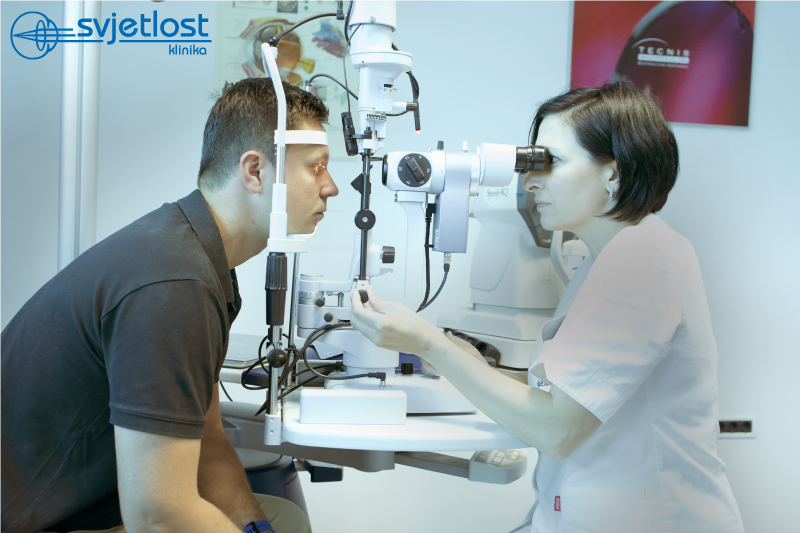
In the latest blog by Maja Pauk Gulic, MD, PhD find about the most common corneal diseases and how to treat them.
You've been to three ophthalmologists and two optometrists in the last year and no one has given you good glasses? Or they were a right fit for 3 months but afterwards, even with them, your dioptre is not accurate? Is the light scatters bothering you, especially at night while driving? You may have a corneal disease called keratoconus, and it is a time for you to undergo detailed diagnostic with a Pentacam showing real corneal topography and it takes just a few seconds to make 100 images of the cornea giving the most precise information about the thickness, shape, and curvature of your cornea.
And what is keratoconus?
Keratoconus is a corneal disease occurring in our area 4-5x more often in young men than in women with most common symptoms of the disease being present in population between the age of 12-25 years. It is a degenerative change, non-inflammatory progressive thinning of the cornea, resulting in a cornea acquiring a conical shape, occurring due to the weakness of the collagen. There is a decline in visual acuity, the diopter is unstable and often changes while the light is being a burden more than usual.
Keratoconus is treated in several ways depending on the progressive forms of the disease; the prescription glasses or better still semi-hard and hard lenses are worn in the initial stages. For progressive forms of keratoconus, the method of “corneal fixation” or “freezing” attempt at the stage where the cornea is currently located is treated by the crosslinking method, which is a corneal stabilization method using vitamin B-riboflavin and ultraviolet light. This strengthens the collagen fibers a form of ‘cement’ that slows down the progress of the disease. In the most severe cases where the cornea is blurred and vision is less than 30%, corneal transplantation is indicated.
You got something in your eye, you washed it off, but it still blisters, blushes and bothers you…
Or did you wake up in the morning with red-eye feeling intense pain?
Or maybe you were scratching your eye trying to take your lens off and now you're in pain and your vision is blurred? Maybe your baby got a finger in your eye?
Most likely, in all four cases above, the corneal surface was injured and erosion occurred. In a case you didn't know, the cornea is a very rich innervated tissue whose injury can be very uncomfortable and painful. In the case of corneal surface injuries, redness of the eye, tears, scratching and foreign body feel, blurred vision and hypersensitivity to light occurs. Often edema and redness of the eyebrows occur due to irritation. An examination by an ophthalmologist is required to apply antibiotic therapy and cycloplegics to prevent possible infection and its possible complications and to relieve pain. Frequent moisturizing of the eye with artificial tears and gels for lubrication and regeneration of the epithelium is advised. This helps to heal the eye and for 2-3 days the patient will have eye closed, for pain relief and to reduce discomfort. The good thing is that although this is a very uncomfortable condition, it passes relatively quickly because the most corneal layer of the cornea - epithelium has a strong and rapid regeneration power so that almost every erosion heals in two to three days.
5 interesting facts about the cornea
- The cornea is transparent at the front, protective layer of the eye is conical shaped
- The cornea is the only tissue that has no blood vessels
- It has a remarkable ability to regenerate and for surface injuries to heal within 48 hours
- Laser vision correction is performed on the cornea. Corneal transplantation due to the keratoconus is one of the most successful organs and tissue transplantation in medicine in general


