
With the proliferation of average life expectancy, we have more and more patients with diseased macula. Additionally, a modern way of life increasingly leads to diabetes development, and with it, macular disease.
The yellow spot or macula is part of the eye background thanks to which we have precise vision. This allows us to read, observe various details, notice details in nature. The yellow spot’s diameter is 5.5 millimetres. This small part of the eye gives us so many information which is the reason we perceive diseases endangering the yellow spot as a major problem. Nowadays, when life expectancy has significantly proliferated there are more and more patients with diseased yellow spots. Besides this, a modern lifestyle with an increased risk of diabetes leads to macular disease.
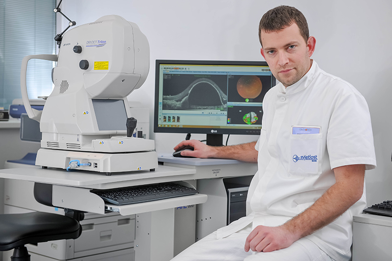
Diagnosis of the yellow spot or macular disease
Macula has a high demand for energy reasons because it has the highest photoreceptors concentration - cells that convert light into electric impulse. This is the reason why it’s most sensitive to metabolic disorders. Even very small changes in the yellow spot can significantly interfere with normal vision. Today, there are modern diagnostic devices that can display even the tiniest details of the yellow spot.
Optical coherent tomography (OCT recording) gives us an insight into the structure of all layers of the yellow spot. The recording process is short and painless. By comparing images before and after therapeutic procedures we can easily see therapy success.
One of the most recent diagnostic methods is OCT angiography - the display of small blood vessels in the yellow spot without the need for intravenous contrast in the circulation. Using this method, all information about the state of microcirculation in the very yellow spot is available in a few seconds. Namely, in the case of diabetes, small blood vessels are killed and thus photoreceptors in yellow spots also die out, which weakens visual acuity.
Anti-VEGF injection therapy
We could only observe the yellow spot in the past. There was no particularly effective treatment method. Nowadays, we have many more therapeutic options. These are primarily anti-VEGF (anti-vascular endothelial growth factor) drugs, present for more than 10 years. With the anti-VEGF injections (Eylea, Lucentis, Avastin) usage in the eye, significant improvement in visual acuity can be achieved both with age-related degeneration of the yellow spot and with complications related to diabetes and other retinal vascular diseases (e.g., central retinal vein thrombosis).
Laser treatment of the yellow spot
Laser therapy can also be applied for the vascular diseases of the yellow spot. Unlike the former conventional laser, there is much lighter laser today, leaving no scars and ideal for yellow spot treatment - so-called endpoint management. Recently, At the Svjetlost Eye Clinic, we are using the newest technology, state-of-the-art for retinal photocoagulation, Pascal Synthesis. The advantage of Pascal laser lies under greater precision and efficiency where laser allows much more “stamps/seals” in a shorter time interval with therapy being significantly less painful. Also, fewer treatments and arrivals to the clinic are accomplished with it.
Eye background surgery - vitrectomy
Certain macular diseases occur as a result of anatomical changes, having a weaker function of the yellow spot (which patient perceives as visual acuity weakening) as a consequence. Such diseases are treated with an operative procedure called vitrectomy. In the case of membranes on yellow spot, ruptures or traction, the only way is to surgically remove membranes and to create optimal anatomical relationships in order to recover macular function.
dr. Marko Vlašić, spec. ophthalmologist, Department of Retina Svjetlost Clinic