




Pediatric Eye Examination
My child needs an eye examination.
01
Pediatric eye examination
A pediatric ophthalmological examination is one of the most important examinations recommended to be performed by the age of four.
A newborn does not have developed vision, which is why the first years of life are crucial for its intensive development. This process lasts until approximately the age of six, and it is never too early to bring a child for an ophthalmological examination.
During the period of visual development, it is important to timely recognize and treat possible disorders to ensure the proper development of the visual system.
Early detection of problems and appropriate treatment can prevent permanent consequences and enable normal visual development.
The examination is adapted to the child’s age and is completely painless.
When treatment is necessary
The most common conditions requiring timely diagnosis and treatment are:
Strabismus
Amblyopia (lazy eye)
Pediatric cataract
Nystagmus
Refractive errors (myopia, farsightedness, astigmatism)
02
What does the examination include?
A pediatric ophthalmological examination lasts up to two hours and includes:
Determination of visual acuity at near and distance
Tests for strabismus and stereovision
Pupil dilation and determination of objective refractive error
Complete examination of the anterior and posterior segments of the eye
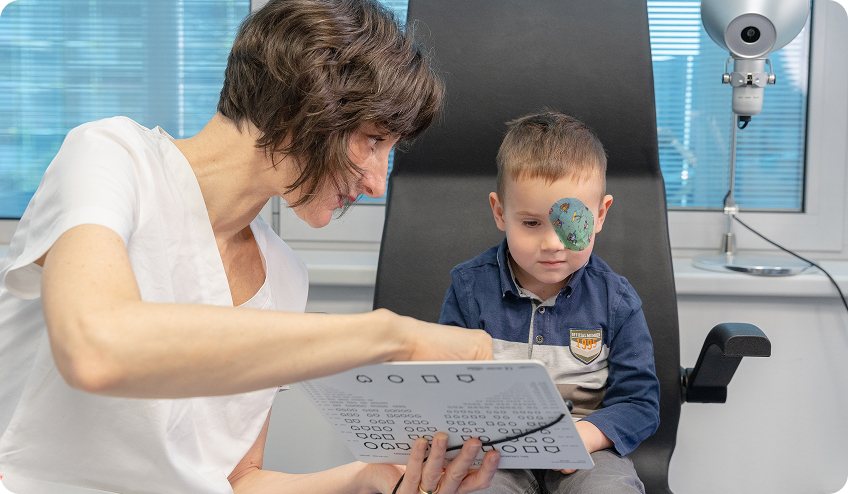
Methods for determining visual acuity differ depending on the child’s age. For infants, special pattern cards are used to assess which pattern size attracts the child’s attention, and the results are then compared with normative tables for their age.
For preschool children, LEA symbols are used, which are pictures the same size as the letters used for adults. To obtain the most precise insight into visual acuity, the child is tested for both near and distance vision. For school-aged children, standard letters are used, as with adults.
Taking an orthoptic status, i.e., strabismus diagnostics, is an important part of the examination for children, and it is also mandatory for adults with strabismus. It serves to assess the position and mobility of the eyes and their cooperation. During this part of the examination, disorders of binocular and stereoscopic vision are identified, meaning it is assessed whether 3D vision exists and if both eyes are working together. The type and magnitude of strabismus, or the angle of deviation, are also determined, and amblyopia and the presence of nystagmus, or eye oscillation, are evaluated. For individuals with nystagmus, additional specific measurements are performed.
Determining objective refraction in children differs from the procedure in adults due to pronounced accommodation, by which a child’s eye can mask even significant refractive errors. To exclude this adaptation, it is necessary to pharmacologically dilate the pupils, which is achieved by repeated instillation of drops, and the entire procedure takes about an hour. Parents are advised to prepare the child in advance for the application of drops and for temporarily blurred vision, which may last several hours, and sometimes until the end of the day.
After complete pupil dilation, objective refraction, or retinoscopy, is performed. This is crucial for detecting hidden refractive errors or other vision disorders and for prescribing the right glasses. Although subjective visual acuity determination requires the child’s cooperation, objective refraction can be determined even in very young children, as it does not depend on their cooperation.
For objective refraction measurement in children, a specially adapted pediatric refractometer, Plusoptix, is also used.
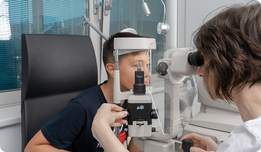
The anterior segment of the eye is examined with a slit lamp microscope to rule out signs of inflammation, allergies, or anatomical abnormalities. Examination of the posterior segment of the eye allows for a detailed assessment of the retina’s appearance.
03
Myopia (nearsightedness) examination
Myopia, or nearsightedness, is a refractive error of the eye where distant objects appear blurry, while close objects are seen clearly.
The most common cause is an elongated eyeball, which causes light rays to focus in front of the retina instead of on it. In recent decades, there has been a significant increase in myopia worldwide, which is associated with prolonged near work, increased use of mobile phones and computers, and insufficient exposure to daylight.
As a child grows, the eyeball also lengthens, meaning its axial length increases, so it is expected that once myopia appears, it will progress over time. Today, methods are available that attempt to slow down this process.
To prevent the development of myopia, it is recommended to spend as much time as possible in daylight, at least two hours a day. Sunlight stimulates dopamine secretion, which is scientifically linked to reducing eye growth in length and slowing down the development of myopia. It is also important to limit screen time, ensure good lighting during reading and near work, and take regular breaks during studying and using digital devices.
When myopia appears, myopia control glasses are prescribed, which differ from conventional glasses. The central part of the lens has a clear vision zone at the correct prescription, while the peripheral part has a different power that creates myopic defocus. This optical effect sends a signal to the eye to slow down its growth in length, thereby slowing the progression of nearsightedness.
If, despite these measures, the refractive error and axial length of the eye continue to progress, low-concentration atropine eye drops may be introduced. These also work to slow down the eye’s growth in length and thus reduce the progression of myopia. They are applied once in the evening before bedtime, and the duration of therapy is individually adjusted to the patient and how the prescription changes. Therapy can last up to two years, and in some cases, even longer.
Timely measures can slow down eye elongation and long-term reduce the risk of complications. In children with more pronounced myopia and an elongated eyeball, the internal structures of the eye, such as the retina, macula, and optic nerve, may be slightly stretched. This increases the risk of more serious problems in adulthood, such as retinal tear or detachment, earlier cataract, and an increased risk of glaucoma. The goal of slowing down nearsightedness is not only better vision in the present but also preserving eye health in the future.
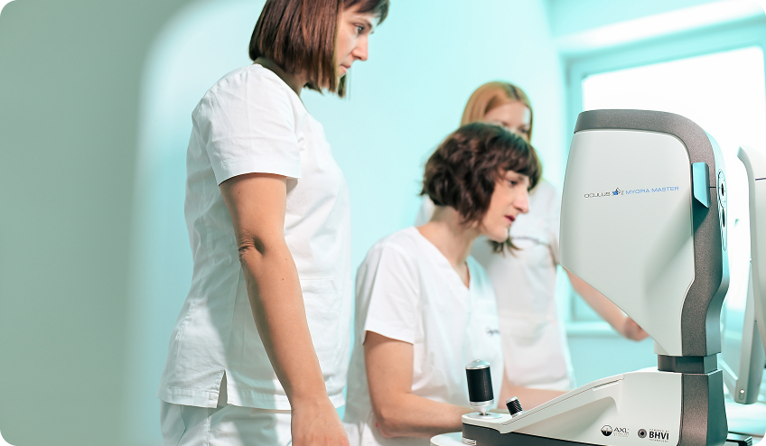
The myopia examination includes a pediatric ophthalmological examination with orthoptic status assessment, visual acuity check, and refraction under mydriasis, along with additional diagnostic tests that allow monitoring of myopia progression. Special devices measure the axial length of the eye and corneal curvature. The scans are short and completely painless. After diagnosis, regular check-ups are recommended every six months, which include prescription checks, axial length measurement, and assessment of treatment effectiveness.
04
Vision condition and treatment
Amblyopia (Lazy Eye)
Amblyopia, also known as lazy eye, is a condition in which visual acuity is reduced in one eye, even though it may appear anatomically normal. If visual acuity is reduced in both eyes, the problem is usually noticed quickly because the child shows clear signs of poorer vision, and parents seek help. However, when amblyopia is present in only one eye, it often goes unnoticed because the child unconsciously relies on the better eye.
In such a situation, the brain uses information from the better eye, while suppressing images from the weaker eye because it cannot combine them into a single perception. If this occurs at an early age, when the visual system is still developing, the weaker eye remains neglected and does not get the opportunity for normal development, which can lead to permanently reduced visual acuity. The risk of developing amblyopia is higher if there is a similar family history of the problem.
Treatment of Amblyopia
Amblyopia treatment begins with the right glasses, followed by occlusion therapy, which involves covering the healthy eye to encourage the weaker eye to work and develop. The duration of occlusion depends on the degree of amblyopia and the child’s age, and can range from one hour to six hours per day.
The patch, or occluder, is placed directly on the skin over the eye, not on glasses, to prevent peeking. It has been proven that the effect of therapy is greater when the child performs near activities during occlusion, such as drawing, reading, or playing. Since children often find it difficult to accept wearing patches, patience and consistency are needed to achieve good results. The more poorly developed the amblyopic eye, the greater the resistance may be, which is an additional reason for persistence in therapy.
The earlier amblyopia is detected, the better the chances of improving vision. In early childhood, the visual system and visual centers in the brain are still plastic enough to compensate for missed development and enable better functional outcomes.
Strabismus
Strabismus is a disorder of eye position or mobility where both eyes do not look in the same direction. In healthy eyes, movements are coordinated, and both eyes are directed towards the same point.
In children, strabismus is most often congenital or occurs at an early age, while in adults it can be a consequence of neurological or systemic diseases affecting eye position or mobility.
Most commonly, the eye deviates inward (esotropia) or outward (exotropia), while upward or downward deviations are rarer. Strabismus is often associated with other vision disorders, such as amblyopia, refractive errors, and nystagmus.
In some cases, eye position can be corrected by wearing appropriate glasses. When this is not sufficient, surgery corrects the eye position and eliminates disturbances such as double vision or asthenopic symptoms, such as fatigue, headaches, and strain. The procedure is intended for both children and adults and contributes to greater safety in daily functioning.
Strabismus in children can hinder proper visual development, which is why it is important to start treatment as early as possible. It is often associated with other vision disorders, and therapy is carried out in combination with other methods. The most common consequence is the development of amblyopia, or lazy eye, in the eye that is not in the correct position. Since amblyopia can only be successfully treated in childhood, it is important to start therapy as early as possible, which includes occluding the healthy eye.
In some forms of strabismus, such as intermittent exotropia, a child may have normal visual acuity and preserved stereopsis. In such cases, regular monitoring is necessary, and if a deterioration of stereopsis is observed, i.e., a weaker ability to recognize stereotests compared to before, surgery may be indicated to preserve or improve stereopsis. In other forms, such as esotropia, surgical treatment is often recommended earlier. The optimal time and type of procedure depend on the type of strabismus.
Each eye is moved by six extraocular muscles, and the impulses for their movement come from the brain and are normally perfectly coordinated for both eyes. This coordination enables binocular vision, i.e., the simultaneous use of both eyes and the creation of a single image. The brain then combines two slightly different images into one three-dimensional perception, which enables stereopsis.
If the eyes are not properly aligned or the mobility of one eye is limited, double vision may occur. In adults, this often leads to the need to close one eye to facilitate daily functioning. In children, due to the greater plasticity of the brain, it can suppress the image from one eye, which increases the risk of amblyopia development. The consequence of this can be the absence of binocular and stereoscopic vision development and permanently reduced function of the affected eye.
Surgical Procedure
If surgery is the best option, after detailed preoperative workup and precise measurements, the procedure is performed on the extraocular muscles that move the eye.
Strabismus surgery is performed under general anesthesia and, depending on the complexity of the procedure, lasts from 30 to 90 minutes.
The child comes for surgery on an empty stomach, with the necessary laboratory results for anesthesia. Before the procedure, the anesthesiologist discusses the child’s general health, previous illnesses, and reviews the results with the parents, after which readiness for surgery is confirmed.
The goal of the surgery is to place the eye in the correct position. This is achieved by strengthening or weakening the extraocular muscles, which is done by repositioning their attachment, shortening, or splitting the muscles.
In cases of muscle paralysis, or reduced mobility, its function cannot be restored, but the eye can be brought into a more favorable position by relocating the weakened muscle or, in some cases, by adjusting the action of healthy muscles to enable movement in the desired direction. Strabismus surgery achieves better alignment of eye positions and their more coordinated joint movement.
Recovery
After strabismus surgery, most patients go home the same day, while if necessary, especially in children, a short stay at the clinic until the first follow-up may be recommended. In that case, a parent can stay with the child in the hospital room during postoperative recovery. In most cases, children leave the clinic a few hours after the procedure.
The first follow-up is performed the day after surgery, while further follow-ups are scheduled depending on the type of procedure, usually at intervals of one to two weeks. Typically, the second follow-up is performed after one week, and the third one month after surgery.
In the early postoperative period, which lasts up to seven days, discomfort, eye redness, and light sensitivity are common. It takes about two weeks for the wound to fully heal and for the eye to become completely white again, and for some patients, it may take up to a month.
After the procedure, the child wears a patch until the first check-up, and it is important to prevent its removal and rubbing of the eyes for a successful recovery. For two to three weeks after surgery, it is necessary to regularly apply the prescribed eye drop therapy. For children, it is especially important to pay attention to the correct application of therapy, as children often reflexively close their eyelids when drops are instilled.
Children usually return to kindergarten or school after two weeks.
Congenital Cataract
Sometimes a child may be born with a cataract in one or both eyes. Congenital cataracts most often result from developmental disorders of the eye, infections during pregnancy, or are genetically determined. When the lens is completely clouded and a clear image does not reach the retina, it is necessary to remove the clouded lens as early as possible to enable proper visual development in that eye.
If the procedure is performed later, there is a significant risk of developing severe amblyopia because the eye did not receive a clear image during the critical period of development and could not develop properly, so surgery must be performed as early as possible. If the clouding is minor or located outside the visual axis, it can be observed during examination, and the image still partially passes to the retina alongside the cataract. In such cases, glasses may be prescribed and occlusion of the healthy eye performed, with regular monitoring.
Congenital Cataract Surgery
During the surgery, a small incision of a few millimeters is made, through which the clouded lens is removed with the help of ultrasound. An artificial lens is implanted in its place, which remains permanently in the eye, is imperceptible, and functionally replaces the natural lens.
After cataract surgery in childhood, treatment must continue, including wearing glasses and occlusion therapy, with the aim of optimal visual acuity development in the operated eye. The procedure is performed under general anesthesia and lasts up to ten minutes per eye. Since a child’s lens is softer than an adult’s, less ultrasonic energy is required, making the procedure less traumatic for the eye.
05
Your medical team – our physicians


Ophthalmologist

Ophthalmologist

Ophthalmology Resident

Ophthalmology Resident
06