Technology
LASER VISION CORRECTION
The Svjetlost Eye Clinic has the Schwind eye-tech-solutions ATOS laser, the Schwind Amaris 1050RS, and performs all methods of laser eye surgery, including All-Laser LASIK and lenticule extraction (SmartSight method).
Svjetlost was among the first clinics in the world to offer third-generation laser vision correction – the SmartSight® method.
Laser technology
Schwind Atos
Schwind ATOS is a femtosecond laser that enables minimally invasive lenticule extraction, as well as the creation of flaps in FemtoLASIK procedures; it is characterized by a very high pulse frequency (up to 4 MHz) and CenTrax technology that automatically compensates eye rotation with active eye tracking, which shortens the duration of the procedure and increases precision.
Schwind Amaris
Schwind AMARIS 1050RS is the most powerful excimer laser in the Amaris family, with an operating frequency of 1050 Hz and an ablation time of only 1.3 seconds per diopter, which increases patient safety and comfort during the procedure.
LASER VISION CORRECTION - DIAGNOSTIC DEVICES
The following are used during the laser vision correction examination:
Corneal topographers
OCULUS Pentacam HR and CSO Sirius – corneal topographers record the relief of the cornea and are the most important tests when selecting candidates for laser vision correction.
In digital form, they show both the actual relief of the cornea and mathematical models that indicate the (non)existence of deviations from a perfectly healthy eye.
In addition to corneal relief, both topographers automatically map higher-order corneal aberrations and display and analyze an image of the entire anterior segment of the eye (anterior chamber, pupil, iris, lens density).
- The OCULUS Pentacam HR is considered the gold standard (reference device), a corneal tomograph that uses Scheimpflug technology to capture 100 corneal images in just two seconds, measuring both the anterior and posterior surfaces of the cornea independently of the tear film, with a curvature measurement accuracy of ±0.1 D and 138,000 processed points in just 2 seconds.
- CSO Sirius combines Placido disk topography with Scheimpflug tomography of the anterior segment of the eye; it measures the pachymetry, elevation, curvature and power of the cornea at a diameter of 12 mm, and also analyzes the pupil size in scotopic, mesopic and photopic lighting conditions.
Aberrometers
The Luneau Visionix Aberrometer and CSO IRX Aberrometer map lower-order (refractive) and higher-order aberrations of the entire eye.
Data on aberrations is obtained from the analysis of over 1500 points.
Objective refraction data eliminate the possibility of incorrect correction and reduce the patient’s need to make a decision about the best image quality (the correct prescription). Laser correction of higher-order aberrations primarily improves the quality of night vision (reduces light scattering). The CSO IRX aberrometer is directly connected to the excimer laser and enables the use of personalized treatments.
- The Luneau Visionix VX120+ is a multi-diagnostic unit that combines an autorefractometer, keratometer, corneal topographer, wavefront aberrometer, pachymeter and non-contact tonometer in one device; it uses the Shack-Hartmann method and Placido ring technology with up to 1300 measurement points and an acquisition time of only 0.2 seconds.
- The CSO iRX3 is a total ocular aberrometer that measures lower and higher order aberrations at up to 45,000 measurement points, with a pyramidal sensor that analyzes the wavefront in real time at up to 33 frames per second.
Pupillometer
The CSO pupillometer measures the size of the pupil in all lighting conditions by changing the lighting conditions during the recording. Information about the largest pupil diameter (pupil at night) is important for planning laser procedures so that the treatment zone is wider than the widest pupil, thus preventing light scattering at night.
How is the examination performed?
The patient sits at the device, leans his head against it, and the device automatically records the cornea in corneal topographers, or the aberrometric profile of the entire eye in aberrometers. The examination is painless, the recording takes a few seconds. The examination is not harmful, and the results are immediately obtained in digital form, or printed out on a printer.
CATARACT AND PRESBYOPIA
CATALYS Precision Laser System
CATALYS femtosecond laser (Johnson & Johnson Vision) is used for laser-assisted cataract surgery. It is a femtosecond laser intended for cataract surgery that uses integrated 3D OCT imaging to perform anterior capsulotomy, lens fragmentation and corneal incisions; enables a personalized surgical plan with extremely high capsulotomy accuracy and a minimal rate of complications compared to the conventional technique.
Bausch + Lomb Stellaris Elite
The lens is removed by phacoemulsification – the cloudy lens is broken up with ultrasound and gently removed – on the Bausch + Lomb Stellaris Elite platform, along with implantation of an artificial intraocular lens.
Bausch + Lomb Stellaris Elite is a platform for phacoemulsification of cataracts with Adaptive Fluidics technology that dynamically adjusts the flow and pressure of the fluid to stabilize the anterior chamber and reduce the so-called surge effect, and Waveform ultrasonic modulation, which breaks up the lens using less energy and heat.
Zeiss ARTEVO 800
A digital 3D operating microscope that shows the surgeon a magnified, highly detailed image of the eye during the entire procedure on a large Ultra HD screen.
The Zeiss ARTEVO 800 is a digital 3D operating microscope that, with 25% higher resolution than conventional optical microscopes, reduces the intensity of light directed into the eye by up to 85%, thereby increasing patient comfort during the procedure, while the AdVision function displays surgical guides for aligning toric lenses on the screen in real time without the need for external marking of the eye.
Zeiss OPMI LUMERA 700
Svjetlost Clinic also uses Zeiss LUMERA operating microscopes in its operating rooms – a classic optical platform with integrated OCT technology – which provides the surgeon with an exceptionally clear and contrasting view of the eye during cataract, cornea, glaucoma, and retina procedures.
Zeiss OPMI LUMERA 700 is an operating microscope with patented Stereo Coaxial Illumination (SCI) technology that provides a highly contrasting and stable red reflex, motorized zoom optics with a ratio of 1:6 and apochromatic lenses for precise visualization; integrated spectral OCT (27,000 A-scans per second) and the ZEISS CALLISTO eye system enable automated, markerless alignment of toric lenses, while additional lenses (128D wide-angle, 60D macular) extend the application to retinal and corneal procedures (e.g. DMEK/DALK).
CATARACT AND PRESBYOPIA - DIAGNOSTIC DEVICES
IOLMaster
IOLMaster – a device that measures the power of the lens that will be implanted in the eye during cataract surgery. Taking into account the length of the eye, keratometric values, the depth of the anterior chamber, and a series of formulas, it calculates the power of the lens that will be implanted.
It is performed by the patient sitting at the device, leaning their head against it, and the device automatically records all the necessary data. The examination is painless, lasts half a minute, and the results are printed out immediately.
The Zeiss IOLMaster 700 is an optical biometer with Swept-Source OCT technology that measures the dimensions of the eye and cornea to calculate the power of the artificial lens; the Total Keratometry function directly measures the posterior surface of the cornea, which reduces the number of refractive surprises after cataract surgery, especially in eyes that have previously undergone laser correction.
Ultrasound + A-scan
Ultrasound + A-scan – a test that provides insight into the inside of the eye and the length of the eye. It is performed with the patient sitting comfortably on a chair, and the doctor gently passes the ultrasound probe over the lower and upper eyelids and takes photographs. With an A-scan, the probe must be placed against the cornea of a previously anesthetized eye. The test is painless, not harmful and lasts a few minutes.
Quantel Medical ultrasound systems (the world’s leading manufacturer of ophthalmic ultrasound) provide A and B scans and high-frequency UBM examinations for precise visualization of the inside of the eye, including measuring the length of the eye to calculate lens power.
Corneal topography
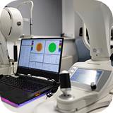
Corneal topography – a test that provides insight into the curvature of the anterior surface of the cornea and determines the value of corneal astigmatism. It is performed by having the patient sit at the device, lean their head against it, and the device automatically records all the necessary data. The test is painless, lasts a few seconds, and the results are printed out immediately.
Endothelial microscope
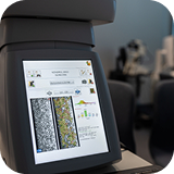
Endothelial microscope – a test that counts the number of endothelial cells in the cornea. It is performed by having the patient sit at the device and rest their chin on the support and the device automatically records all the necessary data. The test is painless, takes a few seconds, and the results are printed out immediately.
OCT of the macula and optic nerve
OCT of the macula and optic nerve – records the structure of the macula and optic nerve and detects possible disorders of these structures, otherwise invisible during examination with a magnifying glass. It is performed by having the patient sit at the device, lean their head against it, and the device automatically records all the necessary data. The examination is painless, lasts a few seconds, and the results are immediately printed out on a printer.
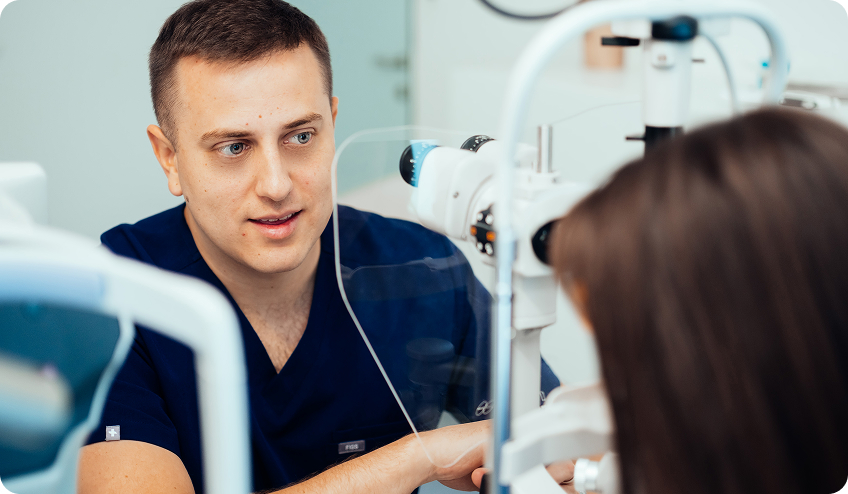
DIABETES AND RETINA
Surgical platform – Stellaris Bausch + Lomb
Platform for minimally invasive vitrectomy through 23-, 25- and 27-gauge ports, with a high cut rate of 10,000 cpm, combined with a unit for cataract surgery with phacoemulsification, two light sources and included laser.
We perform all operations using 3D technology on a large Ultra HD screen, where the surgeon sees the smallest details in the 3D display and can, in effect, look inside the eye.
This is made possible by the 3D Zeiss Artevo 800 microscope .
DIABETES AND RETINA - DIAGNOSTIC DEVICES
OCT – optical coherence tomography (Heidelberg Swept Source OCT + OCT angiography and autofluorescence and Topcon Swept Source OCT)
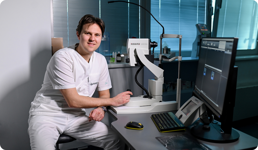
Optical coherence tomography for macula and retinal analysis, with hundreds of thousands of scans per second and a scanning depth that allows for analysis of the retina and choroid. The device also performs OCT angiography, which shows the circulation and the smallest blood vessels of the eye without the use of contrast, and autofluorescence for the detection of retinal dystrophies, as well as degenerative and toxic retinal diseases.
How is the examination performed?
The patient sits at the device, leans his head against it, and the device automatically records the fundus of the eye. The examination is painless, takes only a few seconds, and is not harmful because no radiation is used. The results are obtained immediately, printed on a printer. The examination can also be performed through an undilated pupil.
Fluorescein angiography (Canon 12 MP)
Fluorescein angiography remains a useful test for analyzing circulation in the eye, macular degeneration, diabetic retinopathy, and other retinal diseases. Although today it has largely been replaced by OCT angiography, in some cases it is still irreplaceable and can be crucial in establishing a diagnosis.
How is the examination performed?
The patient sits at the device and rests their chin on the support. A dye is injected into a vein in your arm and reaches the eye in about ten seconds. After which the retinas of both eyes are photographed over a few minutes. The examination is short and the discomfort is minimal. After the contrast agent is administered, your skin may look slightly yellow for about two hours afterwards. The examination results are ready immediately, and the images can be printed on a printer.
Our camera has a high resolution of 12 megapixels and enables retinal imaging in autofluorescence mode, which can help detect some rare retinal diseases.
Eye ultrasound – Quantel Medical
An eye ultrasound is a valuable examination that provides insight into the interior of the eye when the optical media are not transparent, such as in cataracts or hemorrhage. It also complements the clinical findings and helps confirm the diagnosis of numerous retinal diseases. It is most often used in the diagnosis of bleeding in the eye, eye injuries, suspected foreign bodies, retinal detachments, and tumors of the eye and orbit.
How is the examination performed?
The doctor gently passes an ultrasound probe over the lower and upper eyelids and records images of the eye. The examination is painless, non-invasive and only takes a few minutes.
Multifocal electroretinography (mfERG) and electrooculography (EOG)
Devices for recording electrical potentials of the retina enable the detection of numerous hereditary and acquired diseases of the rods and cones of the retina. The examinations are performed simply and painlessly, by placing microelectrodes on the outer part of the eye, in a similar way to recording the electrical activity of the heart or brain.
IRIDEX micropulse multispot laser
The IRIDEX micropulse multispot laser is used for classic retinal photocoagulation, as well as for micropulse laser stimulation of the retina. In classical photocoagulation, it is used to treat diabetic retinopathy and retinal tears. It enables the procedure to be performed faster because it can place dozens of laser spots per second, where older lasers placed one at a time. This makes the procedure faster and more pleasant for the patient.
The procedure is performed on an outpatient basis and only takes a few minutes. The micropulse laser is practically imperceptible and is used to stimulate the retina, for example in diabetic edema and central serous retinopathy. This procedure is also performed on an outpatient basis and only takes a few minutes.
GLAUCOMA
Diagnostic methods and equipment
Measurement of eye pressure: performed by various methods, including classic Goldmann applanation, non-contact tonometry, Pascal tonometry, and iCare handheld portable tonometer. Accurate monitoring of eye pressure is essential for early detection and control of glaucoma.
Computerized visual field testing (perimetry), OCTOPUS 900 – is a device of the latest generation that enables reliable detection of changes in the field of vision caused by various pathological conditions. The examination is non-invasive, painless and performed in a dark room.
- The Haag-Streit OCTOPUS 900 , also described as the “modern Goldmann perimeter”, offers a wide range of static and semi-automated kinetic visual field examinations with guided examination flow and automatic fixation control.
Pachymetry (corneal thickness measurement) – a painless test that measures the thickness of the cornea. A thicker cornea can give a falsely higher measured intraocular pressure: the reading appears elevated, but glaucoma is not present. A thinner cornea gives a falsely lower intraocular pressure value, and is also an independent risk factor. Therefore, corneal thickness is part of the basic workup for glaucoma.
Optical coherence tomography, OCT Topcon Swept Source – is one of the most precise devices of its generation for quick and detailed detection of optic nerve damage and macular degeneration. It enables highly precise analysis of eye structures and helps in early diagnosis and monitoring of diseases.
- The Topcon DRI OCT Triton was the first device in the world to combine Swept-Source OCT technology with multimodal fundus imaging; with a scanning speed of 100,000 A-scans per second, it enables rapid and detailed analysis of the optic nerve and macula.
CHILDREN'S EYE EXAMINATIONS
The Plusoptix children’s binocular autorefractometer measures refraction objectively in young children. It is specially designed and adapted for children from 6 months of age onwards, and is also used for patients with special needs.
It works on the principle of simultaneous binocular diopter measurement from a distance of one meter. As well as refraction, it also measures the size and distance of the pupils.
The measurement is very simple. The child only needs to look at the device for about 0.8 seconds, during which the device emits light and sound signals that attract attention and enable a quick and reliable measurement.
Visual acuity determination
- Preverbal children: preferential looking tests
- Preschool children: Lea charts for children, Lea charts for examining the crowding phenomenon in low vision
- School children: adult optotypes, Hase C and Lea optotypes with varying degrees of “crowding”, a test for examining the crowding phenomenon
Binocular vision tests
- Lang test
- Titmus Fly stereo test
- Bagolini glasses
Prisms
Diagnostic equipment for determining the angle of strabismus is a crucial preoperative examination, especially before strabismus surgery. Based on the measured angle of strabismus, the amount of correction required is determined, that is, how much the eye muscle needs to be moved or shortened to achieve the desired effect.
Eye biometry
IOLMaster/Myopia Master: Biometry is necessary before cataract surgery, as in adults, but also before some strabismus surgeries for adequate planning of operative measures. Today, it is widely used to monitor myopia, which is an increasingly common problem in children.
Cataract surgery equipment
For children’s cataract surgery, the most modern Alcon Infiniti ultrasound platform is used.
CORNEAL DISEASES AND TRANSPLANTATION
Corneal topographer OCULUS Pentacam HR
OCULUS Pentacam HR – corneal topographer records the relief, i.e. the anterior and posterior surfaces of the cornea. In digital form, they display both the actual corneal relief and mathematical models that indicate the presence/absence of deviations from a perfectly healthy eye. In addition to the corneal relief, both topographers automatically map higher-order corneal aberrations and display and analyze an image of the entire anterior segment of the eye (anterior chamber, pupil, iris, lens density).
The Pentacam HR is considered the gold standard (reference device) for corneal topography and takes 100 corneal images in just a few seconds, with 138,000 processed points in just 2 seconds.
How is the examination performed?
The examination is performed so that the patient sits comfortably directly in front of the machine, places his head in the designated places, and follows the instructions of the person performing the examination.
Is the examination painless?
The scan is completely painless because nothing touches the eye, the only thing that is important is that the patient follows the instructions and looks exactly in the given direction.
How long does the scan take?
The examination only takes a few minutes, and several images of each eye are usually taken for better comparison and analysis.
Optical coherence tomography of the anterior segment of the eye – As-OCT (MS39)
The As-OCT MS39 is a non-contact examination, particularly useful in planning surgical procedures on the anterior segment of the eye as well as monitoring the success of surgery after surgery. The device provides high-resolution images. OCT technology can be used to image the anterior and posterior segments of the eye, but it depends on the wavelength of the light.
OCT is excellent for evaluating the cornea (its thickness, LASIK flaps, incisions, dystrophy, scars, etc.) and for imaging the iris, the anterior chamber angle, and the position of the lenses. The examination is performed in the same way as with Pentacam, is painless, and takes an extremely short time (a few seconds per eye).
LUMENIS LASER
For surgical procedures and laser treatments on the eyelids, we use the Lumenis laser – one of the most respected and widely used laser systems in medical aesthetics and ophthalmology.
The Lumenis Laser, widely used in ophthalmology and medical aesthetics with more than 50 years of experience.
Lumenis lasers enable extremely precise cutting and coagulation of tissue, with minimal thermal damage to surrounding structures. This precision makes procedures safer, bleeding is minimized, and recovery is significantly faster than with conventional surgical methods.
In the field of eyelid surgery, the Lumenis laser is used in laser blepharoplasty, the precise removal of changes in the eyelid skin and surrounding area, and post-procedure skin treatment. By combining surgical precision and advanced laser technology, we achieve optimal functional and aesthetic results with a high degree of patient safety.
CONFOCAL MICROSCOPE
Confocal microscope – Heidelberg (HRT3 with Rostock Cornea Module)
Confocal microscopy is an examination that uses laser scanning to image the cornea, conjunctiva, or limbus at the cellular level – a kind of “in vivo histology” of the eye without the need for a biopsy.
The device displays all layers of the cornea (epithelium, Bowman’s membrane, stroma with keratocytes, endothelium), as well as the subbasal corneal nerve plexus. It is used in the diagnosis of corneal inflammation and infection (including acanthamoeba and fungal keratitis), corneal dystrophy, monitoring dry eye, pre- and post-surgical evaluation of the cornea (including transplantation), and in the monitoring of diabetic neuropathy through the analysis of corneal nerve fibers.
Heidelberg HRT3 with Rostock Cornea Module is a compact device that uses confocal scanning laser microscopy to image the cornea, conjunctiva, and limbus at the cellular level, in a field of view of up to 400 × 400 μm, enabling “in vivo histology” – an examination of the corneal structure (epithelium, keratocytes, endothelium, and subbasal nerve plexus) without invasive intervention, useful in the diagnosis of keratitis, corneal dystrophy, dry eye, and diabetic neuropathy.
TOPCON TERA
Topcon TERA is a dry eye imager that brings the entire ocular surface workup together in a single device, non-invasive tear break-up time (NIBUT), tear meniscus height, lipid layer assessment on the Guillon scale, meibography, blink analysis, ocular redness graded on the Efron and Jenvis scales, high-magnification imaging of the eyelid margins, and fluorescein and lissamine green imaging and video; one-touch auto-alignment, focus and capture make every examination faster and more repeatable, so changes in the tear film can be compared reliably from one visit to the next.