



Glaucoma Treatment
I've been told my eye pressure is high.
01
What is glaucoma?
Glaucoma is a progressive disease of the optic nerve that can gradually narrow your field of vision, damage the optic nerve, and cause permanent sight loss that cannot be restored by treatment.
Since the disease often progresses without symptoms, glaucoma is also known as the “silent thief of sight.”
The main risk factor for the development of glaucoma is elevated intraocular pressure, although its value alone does not necessarily indicate the presence of the disease. Normal intraocular pressure values are 10-21 mmHg, but glaucoma can develop even with normal values if there is optic nerve damage and changes in the visual field.
Therefore, glaucoma is considered a complex disease that cannot be diagnosed based on a single finding, but rather by evaluating multiple clinical indicators.
Glaucoma is one of the leading causes of blindness worldwide.
It is estimated to affect 2-3% of individuals over 40 years of age, with a large number of cases remaining undetected. Early detection and regular check-ups are crucial for slowing down the disease and preserving vision.
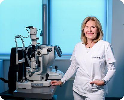
Prof. Smiljka Popović Suić, MD, PhD
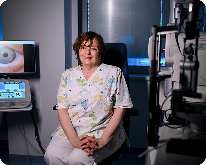
Morena Gavrić, MD
There are several forms of glaucoma.
The most common is open-angle glaucoma, which occurs due to a gradual slowing of the outflow of aqueous humor. This causes fluid to remain in the eye longer than it should, which can lead to elevated intraocular pressure and optic nerve damage.
In angle-closure glaucoma, the cause lies in a drainage angle that is anatomically narrow or completely closed, which significantly restricts the outflow of aqueous humor.
Infantile glaucoma occurs in childhood, while secondary glaucoma develops as a consequence of other conditions, such as eye injuries, inflammation, long-term corticosteroid use, or other eye diseases.
For glaucoma treatment, painless outpatient laser procedures are available, including YAG laser iridotomy, selective laser trabeculoplasty (SLT), and pattern laser trabeculoplasty (PLT), performed with the modern Pascal laser. Surgical treatment includes trabeculectomy and the implantation of an Ex-PRESS mini glaucoma shunt for aqueous humor drainage and lowering intraocular pressure.
These procedures help reduce intraocular pressure and prevent further optic nerve damage, thereby slowing or stopping disease progression and preserving vision.
02
Is this procedure for me?
In its early stages, glaucoma most often causes no symptoms, which is why the disease is often detected only after optic nerve damage has occurred.
Open-angle glaucoma usually progresses slowly and without discomfort, while angle-closure glaucoma can cause sudden and severe symptoms, such as intense eye pain, sudden vision loss, headaches, nausea, and vomiting.
Glaucoma treatment depends on the type and stage of the disease and individual eye characteristics. The goal of every form of treatment is to lower intraocular pressure, preserve the optic nerve, and prevent further vision loss. The ophthalmologist makes the decision on the most appropriate treatment method after a detailed examination and diagnostic workup.
The procedure may be a good choice if:
You have elevated intraocular pressure that cannot be sufficiently controlled with drops
Glaucoma is progressing despite regular therapy
You experience side effects or difficulties using eye drops
You want a more long-term solution for controlling intraocular pressure
You have been diagnosed with a form of glaucoma suitable for laser or surgical treatment
You want to slow down the progression of glaucoma
Individuals over 45 years of age have a higher risk of developing glaucoma, especially if there is a family history of the disease.
Additional risk factors include high myopia, elevated blood pressure, diabetes, thyroid disorders, long-term corticosteroid use, previous eye injuries, and certain anatomical peculiarities of the eye.
03
Glaucoma examination
Although glaucoma cannot always be prevented, its progression can be significantly slowed down with timely detection and treatment.
Regular ophthalmological examinations are important for early detection of the disease, especially in individuals with increased risk.
Examinations are recommended every 2-4 years from the age of 40, and if you are over 65, check your eye pressure every one to two years. Individuals with a family history of glaucoma should begin examinations earlier and have them more often. For individuals already diagnosed with glaucoma, regular check-ups are necessary according to the ophthalmologist’s recommendation and adherence to prescribed therapy.
The examination includes:
Visual acuity check and intraocular pressure measurement
Visual field determination
Measurement of corneal thickness (pachymetry)
Examination of the drainage angle (gonioscopy)
Optic nerve analysis using optical coherence tomography (OCT)
Course of the examination
The examination begins with a consultation with the doctor during which information about symptoms, general health status, and any family history of eye diseases is collected. This is followed by a visual acuity check and an eye examination with a biomicroscope.
The next step is visual field analysis. The procedure is completely painless but requires
good concentration and can take longer, depending on patient cooperation.
After the visual field examination, intraocular pressure is measured. Anesthetic drops are used for the procedure to ensure a comfortable and painless examination. The same drops are used during gonioscopy, an examination of the eye angle that helps determine the type of glaucoma.
If necessary, pupils are dilated with drops to allow a detailed examination of the optic nerve and macula. An important part of the diagnosis is also the OCT examination, which enables precise, fast, and non-invasive analysis of the optic nerve and detection of possible damage.
Due to pupil dilation, vision may be temporarily blurred for several hours after the examination, so we recommend that you do not drive yourself home afterwards.
04
Procedure and recovery
After the examination and analysis of all findings, the doctor recommends further treatment. Glaucoma therapy is aimed at lowering intraocular pressure and preserving the optic nerve, and the choice of method depends on the type and stage of the disease. Medical, laser, and surgical methods are used in treatment today, which are safe, effective, and tailored to each patient.
Treatment Methods and Advantages
Depending on the clinical picture, treatment may include:
Anti-glaucoma eye drops
- easy to apply
- effectively lower intraocular pressure
- slow down disease progression
Laser methods
- YAG laser iridotomy – a fast and effective solution for acute angle-closure glaucoma and a standard method for treating and preventing angle-closure glaucoma
- Selective Laser Trabeculoplasty (SLT, Ellex) – a non-invasive and painless method that improves aqueous humor outflow in open-angle glaucoma
- PLT (Pascal) laser trabeculoplasty – a more modern, comfortable, and effective version of SLT
Surgical methods
- Trabeculectomy – creates a new channel for aqueous humor outflow, reduces intraocular pressure, and stabilizes the disease state
- Ex-PRESS mini glaucoma shunt – a drainage implant that allows for a simpler procedure and faster recovery
Recovery
Recovery depends on the type of procedure, but it is generally fast and without major limitations.

Eye Drops
Applied daily as directed by the doctor, the therapy is completely painless and non-invasive.

After Laser Procedures
The procedure is performed on an outpatient basis, under local anesthesia, lasts only a few minutes, and is completely painless. The patient can return to daily activities immediately after the procedure and continue with eye drop therapy as recommended by the doctor.

After Surgical Procedure
The surgery is performed under local anesthesia and lasts 20 to 30 minutes, after which the patient goes home the same day, with regular intraocular pressure checks and adherence to the doctor's instructions for the most successful recovery.
05
Your medical team – our physicians



06