




Diabetes and the Retina
I have diabetes and I am concerned about my vision.
01
Retinal diseases
Retinal diseases can seriously threaten vision, and without timely treatment, in some cases, they can lead to permanent vision loss.
Early diagnosis and timely therapy are crucial for preserving vision.
One urgent condition is retinal detachment, which requires prompt surgical intervention.
It occurs when the retina tears and a retinal hole forms. Intraocular fluid enters through the retinal hole and detaches the retina from the back of the eye.
Patients most commonly notice the appearance of a dark curtain in their visual field, and if the detachment affects the macula (the center of vision), sudden and significant vision loss can occur. This is precisely why the speed of treatment plays a crucial role.
The foundation of modern retinal surgery is vitrectomy – a minimally invasive procedure that removes the vitreous of the eye to enable access to the retina. It is used for retinal detachment, advanced diabetic retinopathy, macular diseases such as macular hole and epiretinal membrane (macular cellophane), as well as for certain eye injuries and other complex posterior segment diseases.
For diabetic retinopathy, diabetic macular edema, and the wet form of age-related macular degeneration, the standard treatment is intravitreal anti-VEGF drug injections, which reduce retinal swelling, slow disease progression, and help preserve vision. If necessary, treatment is supplemented with laser therapy or surgical intervention. For the dry form of age-related macular degeneration, specific vitamin supplements, regular check-ups, and disease progression monitoring are recommended.
Thanks to modern diagnostic and therapeutic methods, it is now possible to successfully treat a large number of retinal diseases and, in many cases, preserve or significantly improve vision.
Is this procedure for me?
Retinal treatment and surgery are intended for patients with diseases of the posterior segment of the eye that can threaten or significantly reduce vision and require specialist diagnosis and treatment.
This form of treatment may be necessary if you:
Notice sudden or gradual deterioration of vision
Have distorted or wavy images (metamorphopsia)
Experience dark spots, shadows, or a "curtain" in your visual field
Have been diagnosed with diabetic retinopathy or macular disease
Have suspected or confirmed retinal detachment or retinal tear
Require intravitreal therapy (anti-VEGF) or surgical treatment of the vitreous and retina
What does a retinal examination include?
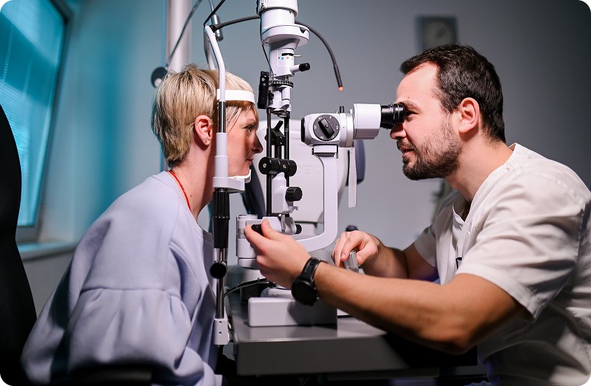
An examination of the back of the eye begins with determining visual acuity in both eyes, followed by intraocular pressure measurement and examination with a biomicroscope to assess corneal clarity and the possible presence of cataracts.
For a detailed examination of the fundus, it is necessary to dilate the pupils with drops. Dilation usually takes between 10 and 30 minutes, and the effect can last about 2 hours. During this period, reading and driving are difficult, so caution is recommended after the examination.
The examination itself is performed in the clinic using a biomicroscope, with the doctor using a lens that is brought close to the eye or gently placed on the anesthetized surface of the eye. This allows detailed examination of the vitreous and retina. The examination is painless and takes approximately one hour. After the examination, brief flashes may occur, which quickly subside.
Stefan Mladenovski, MD
Borivoje Ivezić, MD
What happens after the examination?
After the examination, the doctor makes a diagnosis and determines therapy or recommends additional tests. Diagnostics are supplemented as needed with methods such as OCT (optical coherence tomography) fluorescein angiography or ocular ultrasound. All of these tests can be performed immediately, without additional scheduling.
Based on the findings, further treatment is determined, which may include medications, drops, laser therapy, intravitreal injections, or surgical intervention. In most cases, therapy can be started the same day. In emergency situations, such as retinal detachment or eye injury with a foreign body, surgery can be arranged immediately.
Will a follow-up examination be necessary?
Retinal diseases are often chronic and require regular monitoring. Follow-up examinations are conducted at weekly or monthly intervals, depending on the diagnosis and course of treatment. During follow-ups, pupils may be dilated again and additional tests performed, but such examinations are generally shorter.
02
Retinal detachment and vitrectomy
Retinal detachment is treated with an operation called vitrectomy.
Since the retina is nourished only when it is attached to the back of the eye, any detachment can lead to permanent damage. Therefore, it is important to perform the operation as soon as possible, as delay reduces the possibility of successful vision recovery.
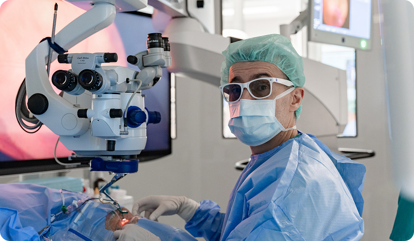
How is the operation performed?
The operation is performed under local anesthesia with sedation. The patient receives a local anesthetic around the eye and sedative medications intravenously, making the procedure completely painless.
During the operation, an anesthesiologist is present who continuously monitors vital functions: blood pressure, heart rate, breathing, and blood oxygen levels.
The procedure takes approximately one hour on average, and most patients go home the same day.
The course of vitrectomy
During vitrectomy, the eye is accessed through three to four very small openings in the sclera, with a diameter of less than 1 millimeter.
Microsurgical instruments are inserted through them to remove:
Diseased vitreous
Blood from the eye
Scar tissue
Pathological blood vessels
Membranes or foreign bodies (depending on findings)
The vitreous is completely removed, and its function is later taken over by natural aqueous humor produced by the eye itself, so the eye continues to function normally.
After removing the vitreous, the surgeon identifies holes in the retina, the retina is reattached, and the damage is sealed with laser treatment.
Completion of the operation – tamponade
At the end of the procedure, a tamponade is placed in the eye – gas, air, or silicone oil – which acts as an “internal bandage” and helps the retina remain attached while it heals.

Gas or air
Spontaneously resorbed within 2-6 weeks, without additional intervention

Silicone oil
Used in more complex cases, remains in the eye for a longer time and is later removed by surgery
In more severe and complex situations, multiple procedures are sometimes necessary to achieve optimal results.
Recovery after vitrectomy
Thanks to modern techniques, this is a minimally invasive procedure without sutures, with minimal stress on the eye and faster recovery. The eye after surgery is usually not very painful or markedly red.
After the procedure, it is necessary to:
Regularly apply prescribed drops and ointment for several weeks
Keep the eye closed on the first day after surgery
Attend follow-up examinations (first day, after 7 days, and after 2-3 weeks)
For about ten days after surgery, rest and careful positioning are needed – usually lying on one side, or sitting with the head down. After that, in uncomplicated detachments, most patients can return to their usual activities. Throughout this time, reading, working on a laptop, or watching television with the other eye is permitted.
The total recovery period is most often between 1 and 4 weeks, although it depends on the type of disease and complexity of the procedure.
Related vision conditions and their treatment
Retinal and vitreous diseases are often interconnected and may require different forms of treatment – from medications and laser therapy to vitrectomy surgery. Below is an overview of the most common conditions and modern treatment methods.

03
Diabetic retinopathy
Diabetes is one of the leading causes of vision impairment and blindness in the working-age population. The eye is particularly sensitive to changes caused by elevated blood sugar, and almost every part of it can be affected. In addition to diabetic retinopathy, inflammation is more common and cataracts appear earlier. The most important ocular complication of diabetes is diabetic retinopathy.
What is diabetic retinopathy?
Diabetic retinopathy is a complication of diabetes in which the retina – the inner layer of the eye responsible for vision – becomes damaged.
The retina converts light stimuli into electrical impulses that travel via the optic nerve to the brain, where an image is formed.
Since diabetes is a disease of small blood vessels (microangiopathy), the retina is particularly sensitive to its damage.
Symptoms of diabetic retinopathy
The main symptom is vision loss, which occurs in two ways:
1. Diabetic macular edema
Elevated sugar levels cause blood vessel permeability in the macular area, leading to fluid accumulation (edema). This fluid damages the sensitive retinal cells and causes a decline in central vision.
2. Proliferative diabetic retinopathy
In long-standing diabetes, blood vessel occlusion and reduced retinal perfusion can occur. In response, new, fragile blood vessels (neovascularization) form. They rupture easily and can cause bleeding in the eye (vitreous hemorrhage), which manifests as sudden blurring of vision or a sensation of “fog” in front of the eye. In advanced cases, scar tissue also develops, which can pull and lift the retina, a serious condition requiring surgical treatment.
Newly formed blood vessels can grow on the iris and in the angle of the anterior chamber, where they obstruct the outflow of aqueous humor and lead to neovascular glaucoma, one of the most severe forms of secondary glaucoma.
Treatment of diabetic retinopathy
Today, the primary form of treatment is intravitreal anti-VEGF therapy, which reduces swelling, bleeding, and prevents the growth of abnormal blood vessels. Corticosteroids, also in the form of intravitreal injections, can be used as a second-line treatment.
Injections are administered under local anesthesia in the form of drops and are painless. Treatment is carried out over multiple sessions, most often over several months or years, depending on the disease status.
Anti-VEGF therapy often produces better results than conventional laser because it can lead to vision improvement, not just stabilization.
If necessary, therapy is supplemented with laser photocoagulation, which further stabilizes the retinal condition. Our clinic uses the modern IRIDEX laser, which enables faster and less uncomfortable treatment, and also has a subthreshold/micropulse laser option for treating the macula itself.
Advanced diabetic retinopathy
If diabetic retinopathy has progressed to a more severe stage of proliferative diabetic retinopathy where newly formed vessels and scars proliferate and bleed into the eye, destroying and detaching the retina, the only treatment is surgery – vitrectomy. Today, given advances in surgical technique and technology, these operations are performed earlier because they are minimally invasive, resulting in better visual outcomes.
Why is early detection important?
In advanced diabetic retinopathy, newly formed vessels and scars destroy healthy retinal tissue either by bleeding or by obstructing healthy circulation. Early treatment with injections and laser achieves destruction of these newly formed blood vessels. Their development can also be prevented if treatment with injections and laser is started in time.
Prevention and regular check-ups
Regular ophthalmological examinations are crucial for all people with diabetes. A check-up is recommended at least once a year, with mandatory pupil dilation and fundus examination, as changes may exist even before symptoms appear.
04
Macular degeneration
Age-related macular degeneration
The macula or yellow spot is the central part of the retina that enables central vision, reading, and recognition of fine details.
Age-related macular degeneration is a disease that most commonly occurs after the age of 50 and leads to gradual loss of central vision. Although peripheral vision remains preserved, reading and facial recognition become difficult.
Risk factors include age, family history, smoking, high blood pressure, and elevated lipids. The disease occurs in two forms: dry and wet.
Dry and wet forms of macular degeneration
The dry form is characterized by gradual degeneration of retinal cells responsible for vision. It progresses more slowly than the wet form, but over time can lead to significant and irreversible loss of visual acuity.
In advanced cases, so-called geographic atrophy occurs, when areas of damaged tissue resembling “gaps” in the retina form in the macula.
For the dry form, there is currently no specific treatment that would completely cure the disease. Vitamin supplements with antioxidants (lutein, zeaxanthin) and omega fatty acids are recommended, which can slow disease progression and transition to the wet form.
The wet form of macular degeneration occurs when pathological blood vessels form beneath the macula that leak fluid and can cause bleeding.
If left untreated, scarring occurs within a period of several weeks to months, which can result in rapid and permanent loss of central vision.
Symptoms of wet macular degeneration
The most common symptoms include:
Central vision loss
Difficulty reading
Dark shadow or missing part of the image in the center of the visual field
Distorted lines (metamorphopsia)
Difficulty distinguishing colors
Slower adaptive vision when transitioning from bright to dark environments
Diagnosis
Diagnosis is made by examining the fundus after pupil dilation, during which the ophthalmologist can detect changes in the macula.
Confirmation of diagnosis is most often done using OCT (optical coherence tomography), and if necessary, fluorescein angiography.
Treatment of wet macular degeneration
Wet macular degeneration is now successfully treated with drugs from the anti-VEGF therapy group (anti-Vascular Endothelial Growth Factor), which reduce fluid leakage and bleeding and can improve vision.
The drugs are administered in the form of intravitreal injections into the eye, under local anesthesia. The procedure is not painful and is performed on an outpatient basis, after which the patient goes home. Treatment is long-term and can last months to years, with repeated injections at regular intervals.
In clinical practice, drugs such as Avastin (bevacizumab), Lucentis (ranibizumab), Eylea (aflibercept), Beovu (brolucizumab), and Vabysmo (faricimab) are used, and new therapeutic approaches are under investigation. All standard therapeutic options for treating wet macular degeneration are available at our clinic.
Why is early detection important?
Regular ophthalmological examinations, especially in older age, enable early detection of macular changes, often even before symptoms appear. Early initiation of anti-VEGF therapy can significantly slow the disease and preserve vision.
05
Epiretinal membrane (macular cellophane)
Diseases of the macular surface
A special group of macular diseases in which a membrane forms over the center of vision are called macular surface diseases and include macular hole, macular cellophane, and vitreomacular traction.
What is a macular hole?
A macular hole is a retinal defect in the macular area and can be a full-thickness tear or involve its layers.
The disease most commonly develops without a clear cause, due to aging of the eye. It is more common in people over 60, and in women. In a smaller number of patients, eye trauma leads to the formation of a macular hole.
Symptoms of macular hole
The disease most commonly develops in one eye and without pain. Since the other eye is usually healthy, symptoms may initially go unnoticed.
The most common symptoms include:
Central vision loss
Difficulty recognizing faces and details
Difficulty reading
Distortion or disappearance of part of letters
Wavy or broken lines
What is epiretinal membrane?
Cellophane maculopathy is a macular disease in which a membrane forms that extends over the center of vision. As the membrane progresses and transitions into denser scar tissue, the disease is referred to as macular pucker.
Symptoms of epiretinal membrane
The most common symptoms are:
Blurred central vision
Image distortion
Wavy lines
Difficulty reading
Diagnosis
Diagnosis is made by examining the fundus after pupil dilation. For precise assessment, OCT (optical coherence tomography) is used, which provides a detailed view of membrane thickness and macular hole size. Assessment of findings is important for treatment decisions, as smaller changes may have mild symptoms, while advanced forms significantly impair vision.
Treatment
Macular hole in most cases requires surgical treatment, while in very early and small changes, a conservative approach can sometimes be considered, although its effectiveness is limited. Cellophane maculopathy is generally treated exclusively surgically.
Vitrectomy for macular diseases
The operation used to treat these diseases is called vitrectomy.
It is a minimally invasive procedure performed under local anesthesia. Through very small openings in the sclera (about 1 mm), the interior of the eye is accessed, where the finest instruments remove changes that pull or cover the macula.
In most cases, the operation achieves hole closure and membrane removal, with a very high success rate. Most patients experience stabilization or improvement of vision, especially if the disease is detected earlier.
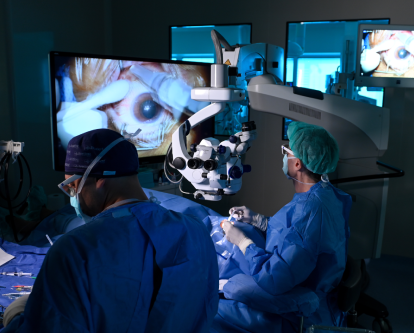
Prof. Ratimir Lazić, MD, PhD
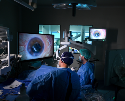
Prof. Ratimir Lazić, MD, PhD
Completion of the operation
At the end of the procedure, air or gas may be placed in the eye, which temporarily supports the retina and macula and helps restore anatomical shape. Gas and air are gradually resorbed over 2 to 4 weeks, so no additional surgical intervention is required.
Recovery
Recovery is generally rapid, and the eye is not markedly painful. Prescribed drops and ointment are used for several weeks. Vision gradually returns as the gas withdraws from the eye. It is possible that the operating surgeon will ask the patient to position themselves lying on their side or sitting with their head down during the first few days.
06
Foreign body inside the eye
A foreign body in the eye (intraocular foreign body) can be of various origins – from dust particles, plant materials, glass to metal.
In more severe cases, it can penetrate the cornea and sclera and damage internal eye structures, including the iris, lens, and retina.
The retina is the deepest layer of the eye that converts light stimuli into nerve impulses, which are then transmitted to the brain where the image we see is formed. Therefore, injuries to deep eye structures represent a serious condition requiring urgent care.
Symptoms of a foreign body in the eye
The most common symptoms include:
Sensation of discomfort or pressure in the eye
Pain and tearing
Photosensitivity
Increased blinking
Inability to open the eye
Redness or bruising of the eye
Superficial foreign bodies most commonly cause corneal scratches/erosions that are successfully treated with drops and therapeutic contact lenses.
If sudden vision loss, fluid or blood leakage from the eye occurs, there is a high risk that it is a foreign body that has penetrated the interior of the eye, and an urgent ophthalmological examination is required.
Such injuries most commonly occur during metalwork (e.g., hammer striking metal).
Eye examination for suspected foreign body
The ophthalmological examination begins with instillation of anesthetic drops to enable detailed examination and possible removal of a superficial foreign body.
The eye is then stained with fluorescein to reveal any corneal scratches and assess the depth of damage.
A mandatory part of the examination is pupil dilation to assess internal eye structures. If necessary, ocular ultrasound is also performed, especially if bleeding or deeper injury is suspected.
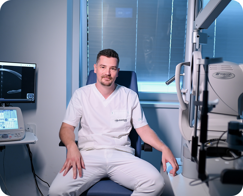
Dino Šabanović, MD
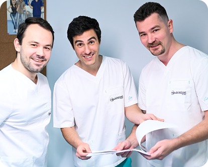
Doctors: Sočo, Ivezić and Šabanović
Treatment of foreign body in the eye
Treatment depends on the depth of injury.
For superficial injuries, antibiotic drops and protective soft contact lenses are used.
If it is a penetrating injury with a foreign body in the eye, the patient undergoes surgical removal of the foreign body and suturing of the cornea or conjunctiva.
If the foreign body is located in the eyeball, deep vitrectomy surgery with retinal management is required. In some cases, traumatic cataract must be treated simultaneously.
The foreign body is removed from the eye using special forceps or magnets. Depending on the size, one of the openings must be enlarged so that the foreign body can be extracted from the eye.
All cases of deep injuries require both local and systemic antibiotic therapy due to the high risk of infection.
Prognosis
Prognosis depends on the degree of damage to internal eye structures and the speed of treatment.
Injuries can lead to corneal clouding, traumatic cataract, or retinal detachment, requiring additional procedures such as corneal transplantation or vitrectomy.
Rapid diagnosis and timely treatment are crucial for preserving vision.
07
Anti-VEGF therapy
Anti-VEGF drugs are a modern group of “smart” drugs that have been used in the treatment of retinal diseases for more than 20 years.
Their use has significantly improved treatment outcomes for previously difficult or incurable retinal diseases.
They are most commonly used in the treatment of:
Wet macular degeneration
Diabetic retinopathy
Retinal vein occlusion
Other retinal diseases associated with edema and newly formed blood vessels
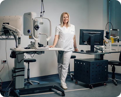
Prof. Nataša Drača, MD, PhD
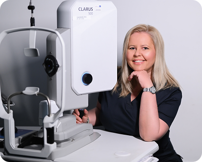
Prof. Nataša Drača, MD, PhD
At Svjetlost Eye Clinic, anti-VEGF therapy has been used since 2005, very soon after its introduction into clinical practice. Today, all available drugs in this class are used, including Avastin, Lucentis, Eylea, Beovu, Eylea 8 mg and Vabysmo.
Svjetlost Eye Clinic takes part in several clinical studies of new anti-VEGF drugs and new ways of administering them. It also holds certifications from regulatory authorities confirming that it meets the highest diagnostic and technological standards for testing new drugs.
How is anti-VEGF therapy administered?
Therapy is conducted on an outpatient basis in a specially prepared sterile environment. The procedure takes about 10-15 minutes and includes:
Local anesthesia with drops
Eye disinfection with iodine
Administration of intravitreal injection with a fine 30-gauge needle
Administration is painless and the patient goes home immediately.
After arriving home, the patient can remove the eye bandage and it is recommended to use artificial tears for several days.
Treatment is administered on a monthly basis and needs to be applied for months or years with possible breaks and regular check-ups.
Role and action of Anti-VEGF drugs
Anti-VEGF drugs work by blocking vascular endothelial growth factor (VEGF), which is responsible for:
Formation of new, pathological blood vessels
Permeability and bleeding
Fluid accumulation in the retina
Thus, they reduce swelling, stop disease progression, and in many cases improve visual acuity.
Fourth-generation anti-VEGF drugs
The latest generation of anti-VEGF drugs works faster and more effectively than earlier drugs and often allows longer intervals between treatments. This means fewer injections and greater convenience for the patient.
It is used in the treatment of:
Wet macular degeneration
Diabetic retinopathy
Central retinal vein occlusion
Compared to earlier drugs, it often allows longer intervals between applications, meaning fewer injections and greater convenience for the patient.
What does this mean for the patient?
Treatment is tailored to each patient, and the number of injections cannot be predicted exactly in advance.
Depending on the type of drug and the severity of the disease, injections are given anywhere from once a month to once or twice a year. Treatment often lasts for months or years, with possible breaks and regular check-ups.
In the initial phase, injections are usually given more often. The intervals are then extended depending on how stable the disease is.
The goal of treatment is to:
Stop disease progression
Stabilize vision
In many cases, improve visual acuity
Regular check-ups are crucial for long-term treatment success.
08
Telescopic lens implantation
Svjetlost Eye Clinic is the first in Croatia and among the first in Europe to perform the surgical procedure of telescopic lens implantation.
Implantable telescopic lens implantation is a modern surgical method intended for people with advanced age-related macular degeneration (AMD) who have experienced significant loss of central vision. In carefully selected patients, the telescopic lens can improve the ability to recognize details and facilitate daily activities such as reading, recognizing faces, or watching television.
Who is the procedure intended for?
Telescopic lens implantation is intended for patients who have:
Advanced age-related macular degeneration (AMD)
Geographic atrophy and marked loss of central vision
Some other diseases that permanently impair vision
Whether a patient is a candidate for the procedure is decided by a retinal specialist after a detailed retinal examination and diagnostic workup.
How does the telescopic lens work?
The implantable telescopic lens contains a special optical system that magnifies the image approximately 2.7 times. The magnified image is projected onto the healthier part of the retina, bypassing the damaged macular area responsible for central vision.
Unlike previously used external telescopic glasses, which have a narrow visual field (up to 20°), SING IMT covers 54° of the central visual field, does not deform facial aesthetics, and frees the user from the need for constant head movements when looking. At the same time, unlike standard intraocular lenses, it is the only option that restores both near and distance vision in severe forms of AMD, while competing devices offer significantly less effect or are limited to only one segment of vision.
Results depend on the degree of eye damage and individual characteristics of each patient.
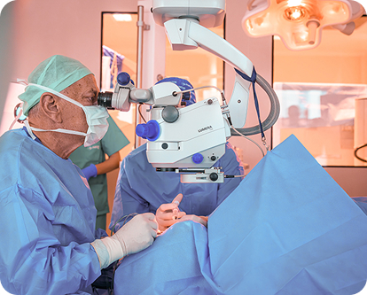
Prof. Nikica Gabrić, MD, PhD
What does the operation look like?
Telescopic lens implantation is performed by microsurgical procedure under local anesthesia. The operation takes approximately 20 to 30 minutes, and during the procedure, the natural eye lens is replaced with an implantable telescopic lens.
The procedure is painless, and most patients go home the same day, with detailed instructions on postoperative treatment and follow-up examinations.
Recovery after the procedure
Recovery does not end with the operation itself. After telescopic lens implantation, a period of adaptation follows during which the eye and brain adjust to the new way of seeing.
An important part of treatment is vision rehabilitation, which includes targeted visual exercises under the guidance of specialists. In most patients, improvement develops gradually over several weeks or months.
Regular check-ups enable monitoring of recovery and adjustment of the rehabilitation program according to individual needs.
What are the advantages of the telescopic lens?
Compared to conventional optical aids, the implantable telescopic lens enables:

Permanent implantation inside the eye
Image magnification without wearing special telescopic glasses
Better utilization of the preserved part of the retina
Improvement of functional vision in selected patients
Greater independence in daily activities
09
Your medical team – our physicians


Ophthalmologist

Ophthalmologist

Ophthalmologist

Ophthalmologist

Ophthalmologist
Ophthalmologist
10