

Patient Examinations
I need an eye examination.
01
Eye examinations and consultations
Eye examinations and consultations enable timely assessment of vision status and monitoring of eye health. Based on the examination, it is possible to accurately determine whether further interventions are necessary and to select the most appropriate approach for each patient.
Examinations also enable early detection of retinal changes, including diabetic damage and degenerative macular diseases, which is crucial for preserving vision.
Assessment of risk and monitoring of elevated intraocular pressure are also performed, which can prevent the development of glaucoma in the early stages of the disease.
In patients who have already undergone surgery, examinations serve to monitor the results of the procedure and provide long-term follow-up of vision stability.
02
General ophthalmological examination
A general ophthalmological examination is a fundamental examination that assesses eye health, determines refractive error, and detects possible eye diseases in their early stages.
What does the examination include?
Determination of visual acuity and refractive error for distance and near vision
Measurement of intraocular pressure
Examination of the anterior segment of the eye using a biomicroscope (eyelids, conjunctiva, cornea, anterior chamber, pupil, and lens)
Pupil dilation and examination of the fundus, including the vitreous, optic nerve, retina, and blood vessels
If the eye is healthy and only refractive error is determined, appropriate vision correction will be recommended (glasses for distance, near vision, or progressive lenses).
If an eye disease is detected during the examination, such as inflammation, cataract, dry eye syndrome, eyelid disease, macular disease, diabetic retinopathy, or glaucoma, the physician will recommend appropriate therapy, additional diagnostic tests, or examination by a subspecialist.
Duration of the examination
The examination takes approximately 60 minutes on average, if no additional tests are required.
Preparation for the examination
It is recommended to bring to the examination:
- previous medical documentation, if available
- existing glasses
If you wear contact lenses, they must be removed before the examination:
- soft contact lenses at least 24 hours before the examination
- hard and semi-rigid contact lenses for as many days as you have worn them in years, and bring them in their case
Glasses may be worn without restriction.
Important information
Due to pupil dilation, vision may be temporarily blurred for several hours after the examination. For this reason, we recommend that you do not drive a motor vehicle after the examination.
03
Contact lens examination
A contact lens examination is designed to select the most suitable contact lenses according to your refractive error, eye structure, and daily needs. The examination is performed by ophthalmologists specialized in prescribing contact lenses.
What does the examination include?
In addition to a general ophthalmological examination, the following are determined during the examination:
Corneal curvature
Tear film quality
Selection and trial of trial contact lenses to find the optimal solution for each patient
At our clinic, we prescribe soft, semi-rigid, and hard contact lenses, as well as special contact lenses for keratoconus.
Duration of the examination
The duration of the examination depends on the type of contact lenses and the individual needs of the patient, and takes approximately 60 minutes on average.
Preparation for the examination
When scheduling, it is necessary to emphasize that you are coming for a contact lens examination. It is also important to mention whether you already wear contact lenses and what type you use (soft, semi-rigid, or hard).
It is recommended to bring to the examination:
- the contact lenses you currently wear
- existing glasses
- previous medical documentation, if available
To ensure the most accurate measurements, it is necessary to temporarily stop wearing contact lenses:
- soft contact lenses at least 24 hours before the examination
- semi-rigid and hard contact lenses for as many days as you have worn them in years
Important information
Due to pupil dilation, vision may be slightly blurred for several hours after the examination, so driving is not recommended during this time.
04
Multifocal lens implantation examination
An examination for multifocal intraocular lens (IOL) implantation represents a detailed diagnostic evaluation of the eye with the aim of selecting the optimal intraocular lens and assessing the patient’s visual potential.
During the examination, the specialist assesses the health status of the eye, visual needs, and patient expectations in order to select the most appropriate intraocular lens. There are several types of lenses, and the final choice depends on the eye findings and individual needs.
What does the examination include?
The examination includes:
Visual acuity determination
Measurement of intraocular pressure
Biomicroscopic examination of the anterior and posterior segments of the eye
Pupil dilation and detailed examination of the fundus
Additional diagnostic tests include:
- IOL Master (calculation of intraocular lens power)
- corneal topography
- OCT of the macula
- ocular ultrasound
- measurement of corneal endothelial cell count
- biomicroscopic examination of the fundus
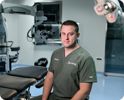
Krešimir Gabrić, MD
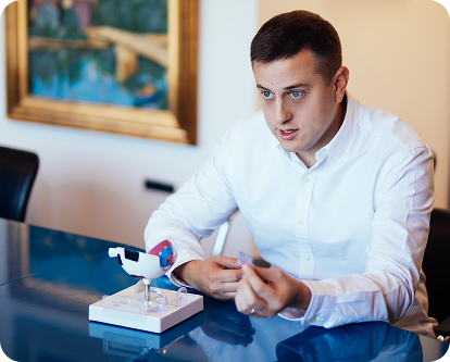
Krešimir Gabrić, MD
Duration of the examination
The examination takes approximately 90 to 120 minutes, depending on the required tests.
Preparation for the examination
It is recommended to bring to the examination:
- previous medical documentation, if available
- existing glasses
If you wear contact lenses:
- soft lenses at least 3 days before the examination
- hard or semi-rigid lenses at least 14 days before the examination
Important information
Due to pupil dilation, vision may be temporarily blurred after the examination. Driving a motor vehicle is not recommended for several hours after the examination.
05
Examination for cataract surgery
During the cataract surgery examination, the condition of the eye, the degree of lens opacity, and detailed diagnostics are assessed for the purpose of planning the surgery and selecting the intraocular lens.
What does the examination include?
The examination includes:
Visual acuity determination
Measurement of intraocular pressure
Biomicroscopic examination of the eye
Pupil dilation and detailed examination of the fundus (lens, optic nerve, macula, and retina)
Diagnostic tests
Preoperative evaluation includes:
IOL Master – precise calculation of intraocular lens power.
Ultrasound A-scan – for dense cataracts, calculates lens power.
Corneal topography – analysis of astigmatism and corneal shape.
OCT of the macula and optic nerve – assessment of macular and optic nerve status.
Ocular ultrasound – for opacities when the fundus cannot be clearly examined.
Measurement of corneal endothelial cells – assessment of corneal health before surgery.
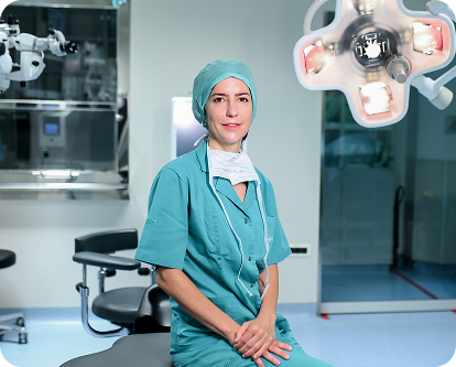
Prof. Maja Bohač, MD, PhD
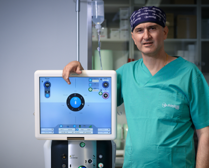
Ante Barišić, MD, PhD
Duration of the examination
The examination takes approximately 90 to 120 minutes, depending on the required tests.
Preparation for the examination
It is recommended to bring to the examination:
- previous medical documentation, if available
- existing glasses
If you wear contact lenses:
- soft lenses at least 3 days before the examination
- hard or semi-rigid lenses at least 14 days before the examination
Important information
Due to pupil dilation, vision may be temporarily blurred after the examination. Driving a motor vehicle is not recommended for several hours after the examination.
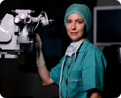
Prof. Maja Bohač, MD, PhD
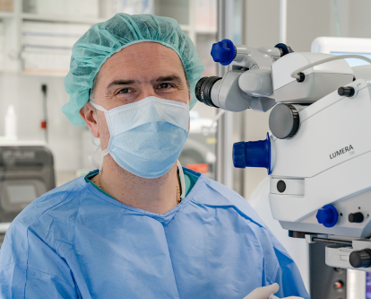
Ante Barišić, MD, PhD
06
Retinal examination
A retinal examination is a detailed examination of the posterior segment of the eye (vitreous, retina, and optic nerve) that diagnoses fundus diseases and assesses vision status.
What does the examination include?
The examination begins with:
Determination of visual acuity in both eyes
Measurement of intraocular pressure
Biomicroscopic examination of the anterior segment of the eye
Pupil dilation with drops
Detailed examination of the fundus (retina and vitreous)
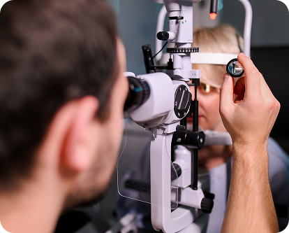
The examination is performed using a biomicroscope, during which the physician uses a lens that is brought close to the eye or gently placed on the anesthetized surface of the eye. This allows detailed examination of the vitreous and retina.
Additional diagnostic tests
If necessary, the examination is supplemented with:
- OCT (optical coherence tomography)
- fluorescein angiography
- ocular ultrasound
Duration of the examination
The examination takes approximately 90 to 120 minutes, depending on the required tests.
Preparation for the examination
It is recommended to bring to the examination:
- previous medical documentation, if available
- existing glasses or contact lens case (if you wear them)
Contact lenses do not need to be removed in advance.
Important information
Due to pupil dilation, vision may be temporarily blurred after the examination. Driving a motor vehicle is not recommended for several hours after the examination.
The examination is painless, and after the examination, brief flashes may occur that resolve spontaneously.
Based on the findings, therapy is determined, which may include drops, medications, laser treatment, intravitreal injections, or surgical intervention. In urgent cases, emergency treatment or surgery on the same day is possible.
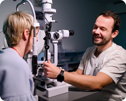
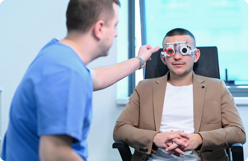
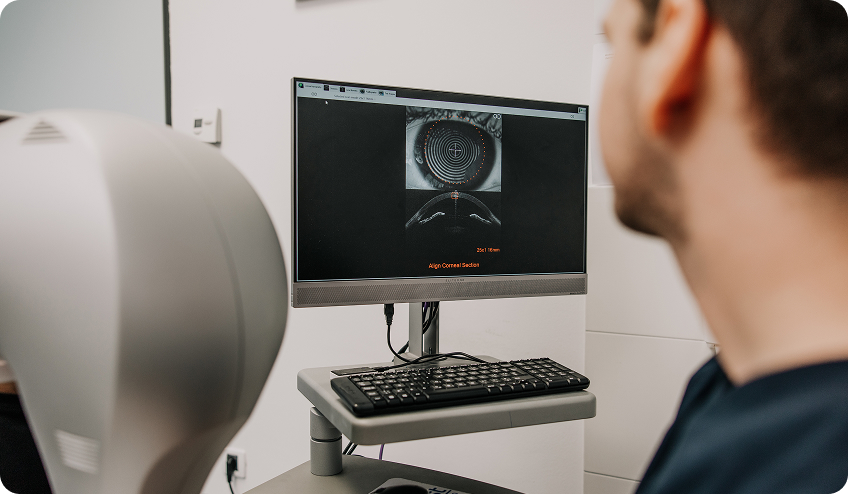
Lovro Sočo, MD during patient examination
Patient examination
07
Glaucoma examination
A glaucoma examination is a detailed diagnostic evaluation that assesses intraocular pressure, optic nerve status, and the risk or presence of glaucoma.
What does the examination include?
The examination includes:
General Ophthalmological Examination
Visual Field
Pachymetry (measurement of corneal thickness)
Gonioscopy (examination of the angle of the eye)
OCT (optical coherence tomography of the retina and optic nerve)
Duration of the examination
The examination may take 2 to 3 hours.
Preparation for the examination
It is recommended to bring to the examination:
- previous medical documentation, if available
- existing glasses
Important information
During part of the examination, anesthetic drops may be applied, so tests such as gonioscopy and intraocular pressure measurement are painless.
Visual field testing requires concentration and may extend the duration of the examination.
Due to pupil dilation, vision may be temporarily blurred after the examination. Driving a motor vehicle is not recommended for several hours after the examination.
08
Corneal transplant examination
A corneal transplant examination includes a detailed preoperative ophthalmological evaluation to assess the condition of the eye and plan the surgical procedure.
What does the examination include?
Detailed examination using a biomicroscope
Visual acuity determination
Measurement of intraocular pressure
Corneal topography
Measurement of corneal thickness (pachymetry)
Assessment of anterior chamber depth
Examination of the fundus
Ocular ultrasound
Biometric measurements and intraocular lens calculation, if combined surgery is planned
Duration of the examination
The examination takes approximately 60 to 90 minutes on average, depending on the required tests.
Preparation for the examination
It is recommended to bring to the examination:
- previous medical documentation
- existing glasses
Important information
Due to pupil dilation, vision may be temporarily blurred after the examination. Driving a motor vehicle is not recommended for several hours after the examination.
09
Keratoconus examination
A keratoconus examination is a detailed ophthalmological examination that assesses the shape, structure, and stability of the cornea and detects or monitors changes characteristic of keratoconus.
What does the examination include?
Detailed examination using a biomicroscope
Visual acuity determination
Measurement of intraocular pressure
Corneal topography
Measurement of corneal thickness (pachymetry)
Assessment of anterior chamber depth
Examination of the fundus
Duration of the examination
The examination takes approximately 60 to 90 minutes on average, depending on the scope of diagnostics and required imaging.
Preparation for the examination
It is recommended to bring to the examination:
- previous medical documentation
- existing glasses/lenses
If you wear contact lenses, they must be removed before the examination:
- soft contact lenses at least 72 hours before the examination
- hard or semi-rigid contact lenses at least 10 days before the examination
Important information
Due to pupil dilation, vision may be temporarily blurred after the examination. Driving a motor vehicle is not recommended for several hours after the examination.
10
Eyelid surgery examination
An eyelid surgery examination includes a detailed ophthalmological and aesthetic-functional assessment of the eyelid area and surrounding eye structures. The aim of the examination is to assess the function, position, and appearance of the eyelids and to plan aesthetic and/or reconstructive surgery.
What does the examination include?
Determination of visual acuity
Determination of refractive error using an automatic refractometer
Measurement of intraocular pressure (tonometry, IOP)
Examination of the anterior segment of the eye using a biomicroscope
Assessment of tear film (Schirmer test)
Pupil dilation and examination of the fundus, including the optic nerve, retina, and center of vision
Objective assessment of refractive error
Assessment of eyelid position and function includes:
Measurement of palpebral fissure height and width
Measurement of eyelid distance from the central light reflex (MRD1 and MRD2)
Assessment of levator muscle function
Assessment of eyelid elasticity
Analysis of eyeball position in relation to the eyelids and surrounding bony structures
Assessment of eye motility and ocular muscle function
Duration of the examination
The examination takes approximately 60 minutes on average.
Preparation for the examination
It is recommended to bring to the examination:
- previous medical documentation
- existing glasses
Important information
Due to pupil dilation, vision may be temporarily blurred after the examination. Driving a motor vehicle is not recommended for several hours after the examination.
11
Strabismus examination
A strabismus examination includes a detailed ophthalmological and orthoptic evaluation with the aim of assessing the position, motility, and coordination of both eyes, as well as determining the type and magnitude of strabismus. The examination is particularly recommended for patients with strabismus and in childhood.
What does the examination include?
Determination of visual acuity
Determination of refractive error using an automatic refractometer
Measurement of intraocular pressure (tonometry, IOP)
Examination of the anterior segment of the eye using a biomicroscope
Assessment of tear film (Schirmer test)
Pupil dilation and examination of the fundus, including the optic nerve, retina, and center of vision
Objective assessment of refractive error
Assessment of strabismus includes:
Determination of the type and magnitude of strabismus
Measurement of the angle of deviation in prism diopters (PD), at near and distance and in all directions of gaze
Assessment of eye motility for each eye separately (ductions)
Assessment of coordinated eye movements (versions and vergences)
Assessment of binocular vision and stereovision (Bagolini, Titmus, Lang tests)
Testing for diplopia (if necessary)
Measurement of accommodation (near adaptation)
Orthoptic status
In case of planned strabismus surgery, a simulation of postoperative eye position using special prisms/lenses may also be performed if necessary.
Duration of the examination
The examination takes approximately 60 to 90 minutes on average, depending on the scope of orthoptic testing.
Preparation for the examination
It is recommended to bring to the examination:
- previous medical documentation
- existing glasses
Important information
Due to pupil dilation, vision may be temporarily blurred after the examination. Driving a motor vehicle is not recommended for several hours after the examination.
12
Pediatric eye examination
A pediatric ophthalmological examination is a detailed examination of vision and eye structures adapted to the child’s age.
What does the examination include?
Determination of visual acuity at near and distance
Tests for strabismus and stereovision
Pupil dilation and determination of objective refractive error
Examination of the anterior and posterior segments of the eye
Methods for determining visual acuity:
For infants: special cards and monitoring of response to patterns
Preschool age: Lea symbols (pictures)
School age: standard letter charts
The orthoptic part of the examination includes:
- assessment of eye position and motility
- determination of strabismus and its magnitude
- assessment of binocular vision (cooperation of both eyes), diagnosis of amblyopia, assessment of nystagmus (additional measurements if necessary)
- stereovision tests (Titmus, Lang)
Assessment of refractive error includes:
- pupil dilation in several stages (duration approximately 60 minutes)
- determination of objective refractive error after pupil dilation (skiascopy)
- measurement with a pediatric autorefractometer (Plusoptix) if necessary
Duration of the examination
The examination takes up to 120 minutes on average, depending on the child’s cooperation and required tests.
Preparation for the examination
It is recommended to bring to the examination:
- previous medical documentation
- existing glasses (if the child wears them)
If the child wears contact lenses, they must be removed before the examination:
- soft contact lenses at least 24 hours before the examination
- lenses must be brought in their case to the examination
Glasses may be worn without any restrictions.
Important information
Due to pupil dilation, the child will have blurred vision after the examination, which may last from several hours to the entire day. It is recommended that the child be prepared in advance for the application of pupil dilation drops.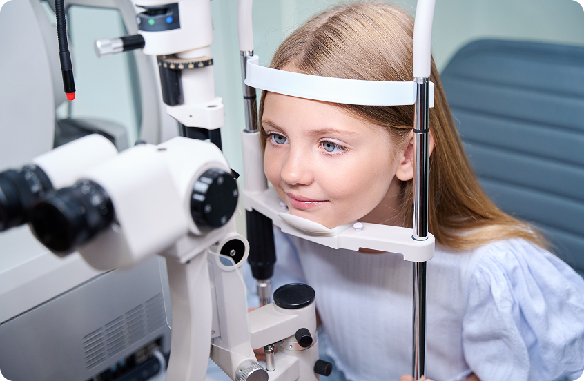
13
Myopia (nearsightedness) examination
Myopia or nearsightedness is a refractive error of the eye in which the child sees distant objects blurred, while near objects are clear. The most common cause is an elongated eyeball, which causes light rays to focus in front of the retina instead of on it. In recent decades, there has been a significant increase in nearsightedness in children worldwide. It is believed that this is due to increased time spent in front of screens, prolonged near work, and less time spent outdoors in daylight.
Can the development of nearsightedness be prevented?
Although it is not always possible to prevent the onset of myopia, certain habits can reduce the risk of its development and slow its progression:
Spend at least 2 hours daily outdoors in daylight
Ensure good lighting during reading and writing
Take regular breaks during studying and near work
Exposure to daylight stimulates dopamine secretion, which has been proven to slow eye growth in length and reduce the risk of developing nearsightedness.
Why is it important to slow the progression of myopia?
In children with myopia, the eyeball gradually elongates, which is why refractive error most often increases during childhood and adolescence.
Greater nearsightedness is associated with an increased risk of developing eye diseases in adulthood, including:
Retinal tears and detachment
Glaucoma
Earlier development of cataract
Macular changes
The goal of treatment is not only to ensure good vision today, but to preserve eye health in the long term.
Myopia control
Today, there are effective methods by which we can slow the progression of nearsightedness.
Myopia control glasses
Myopia control glasses differ from conventional glasses. In myopia control glasses, the center of the lens has a focus area with the appropriate refractive error that enables clear vision, while the peripheral part has a lower refractive error and creates myopic defocus, which sends a signal to the eye to slow growth in length, thereby slowing the progression of myopia.
Atropine drops
If, despite the measures taken, refractive error and axial eye length continue to increase, it is possible to introduce therapy with atropine drops in a highly diluted concentration.
The drops are most often applied once daily before bedtime, and the duration of therapy is individually adapted to each child. Therapy may last several years, depending on the rate of myopia progression.
Myopia examination and monitoring
Examination for assessment and monitoring of nearsightedness includes:
- determination of visual acuity
- orthoptic examination
- determination of refractive error after pupil dilation
- examination of the anterior and posterior segments of the eye
- measurement of axial eye length
- measurement of corneal curvature
All measurements are quick, painless, and adapted to children.
After diagnosis, regular check-ups every 6 months are recommended. At check-ups, we monitor changes in refractive error, eye growth, and the effectiveness of prescribed therapy in order to adjust treatment in a timely manner.
14
Dry eye examination
Disease description
Dry eye syndrome is a common disorder of the ocular surface caused by reduced tear production or impaired tear film quality. The tear film plays a key role in maintaining corneal health, vision stability, and protecting the eye from infections.
In dry eye, tear film instability and increased tear evaporation occur, which can lead to ocular surface damage and chronic inflammation.
The most common symptoms include:
Sensation of dryness and stinging in the eyes
A gritty or foreign body sensation in the eye
Redness of the eyes
Blurred or fluctuating vision
Increased reflex tearing
Sensitivity to light
Eye fatigue, especially when working on a computer or reading
Symptoms are often more pronounced in air-conditioned or heated rooms, during prolonged screen work, and when wearing contact lenses.
Purpose of the examination
The purpose of the examination is to:
Diagnose dry eye syndrome
Classify the type of dry eye
Objectively document findings
Assess the degree of ocular surface damage
Prescribe targeted and individualized therapy
Based on the findings, a treatment and disease monitoring plan is developed.
A follow-up examination is usually planned for 6-8 weeks to assess the effect of therapy and adjust the treatment plan if necessary.
Classification of dry eye
The examination enables differentiation of the main types of dry eye:

Aqueous-deficient dry eye
Caused by reduced lacrimal gland function.

Evaporative dry eye
Most commonly associated with meibomian gland dysfunction (MGD), which produces the lipid layer of the tear film.

Mixed type dry eye
A combination of both mechanisms, which is the most common form in clinical practice.
Course of the examination
The examination consists of several phases:
- completion of the OSDI-6 questionnaire
- diagnostic imaging of the ocular surface
- clinical examination by the physician
- final consultation and treatment plan
The OSDI-6 questionnaire is used to assess:
- symptom intensity
- impact of symptoms on daily activities
- subjective severity of the disease
Instructions before the examination
Bring a list of medications you take regularly
Bring previous ophthalmological and other specialist findings
Do not use eye drops 2-4 hours before the examination (unless the physician has advised otherwise)
Come without contact lenses (remove soft contact lenses at least 24 hours before the examination, and hard and semi-rigid contact lenses for as many days as you have worn them in years)
It is recommended to come accompanied, as after pupil dilation there may be temporary blurred vision and light sensitivity.
Patient Admission
Upon admission, the patient receives the OSDI-6 questionnaire. The patient completes it before the examination begins and submits it to the doctor.
Diagnostic Imaging
Diagnostics are performed according to a standardized dry eye examination protocol.
The examination includes:

Non-invasive TBUT (NIBUT)
Assessment of tear film stability without contact with the eye surface.

Anterior Segment OCT (MS-39)
Corneal epithelial relief map, analysis of epithelial quality, corneal cross-section in two planes, and assessment of tear meniscus height.

Meibography (OFA)
Analysis of meibomian gland structure and assessment of the degree of their atrophy or dysfunction.
If necessary, slit-lamp photodocumentation is also performed to monitor changes on the eye surface.
Clinical Examination
The clinical examination includes:
Visual acuity determination
Tonometry (ICARE)
Eyelid and eyelid margin examination
Tear film quality assessment
Additional diagnostic tests are also performed:

Fluorescein staining of the eye surface
Assessment of corneal and conjunctival epithelial damage. Findings can be classified according to standard scales (Oxford grading)

TBUT test
Assessment of tear film stability. Values shorter than approximately 10 seconds may indicate an unstable tear film.

Schirmer test
Assessment of the amount of tears produced, used to differentiate reduced tear production from evaporative dry eye.

Meibomian gland assessment
Quality of secretion, gland patency, signs of dysfunction (MGD).
This is followed by:
- instillation of drops
- pupil dilation
- fundus examination (fundoscopy)
This assesses the overall condition of the eye and rules out other eye diseases.
Interpretation of findings
Based on the results obtained, the doctor assesses:
- type of dry eye
- tear film stability
- meibomian gland function
- degree of eye surface damage
- disease severity (mild, moderate, or severe form)
All findings are documented in the medical record, along with photodocumentation and measurement results.
Final consultation
At the end of the examination, the doctor:
- explains the findings to the patient
- classifies the type of dry eye
- determines an individualized treatment plan
- provides advice on: eyelid hygiene, proper use of artificial tears, computer work habits, eye surface protection
The patient receives a written report with recommended therapy.
A follow-up examination is usually planned for 6 to 8 weeks later, to assess the response to therapy and adjust the treatment plan if necessary.
15
Second opinion from an ophthalmologist – ophthalmological examination
An ophthalmological examination for a second opinion is intended for patients who want an additional assessment of their findings, diagnosis, or recommended treatment.
During the examination, the ophthalmologist thoroughly analyzes existing medical documentation, previous findings, and symptoms, and performs additional ophthalmological diagnostics as needed.
The goal is to confirm the existing diagnosis, clarify any ambiguities, and assess whether other treatment options exist.
What does the examination include?
Discussion of complaints and the course of the disease to date
Review of existing medical documentation and findings
Complete general ophthalmological examination
Assessment of the existing diagnosis and recommended treatment
Additional diagnostic tests or subspecialist evaluation as needed
Explanation of findings and possible options
Recommendation for further action
When is a second opinion useful?
A second opinion can provide additional reassurance and help you make an informed decision about your health.
Doctors strive to provide each patient with a clear and understandable explanation of their condition and to answer all questions related to the diagnosis and treatment options.
16
Corneal confocal microscopy
Corneal confocal microscopy is a non-invasive diagnostic method that provides a detailed view of the cornea at a cellular level. It is used for:
Assessment of corneal nerve fibers and their changes
Diagnosis and monitoring of corneal neuropathy and neuropathic ocular pain
Assessment of dry eye syndrome and ocular surface diseases
Detection and monitoring of infectious keratitis, especially when Acanthamoeba or fungal infections are suspected
Assessment of corneal dystrophies and degenerations
Analysis of endothelium, epithelium, stroma, and inflammatory cells
Monitoring changes after refractive and other corneal surgeries and transplantation
Assessment of changes associated with contact lenses
Research and monitoring of corneal nerve changes in diabetes, glaucoma, and certain neurological and systemic diseases
The examination is fast, minimally invasive, and provides insight into microscopic changes that cannot be seen with a standard ophthalmological examination.
17