



Corneal Diseases
My cornea is damaged and my sight has become cloudy.

01
Which treatment method is right for me?
The treatment of corneal diseases depends on the type and stage of damage, as well as the individual characteristics of the eye. The goal of corneal procedures can be disease stabilization, vision improvement, or restoration of corneal clarity.
The decision on the optimal treatment method, whether it is cross-linking, the Cornea Lux method, the CAIRS method, or corneal transplantation, is made by an ophthalmologist after a detailed examination.
The procedure may be a good choice if:
- You have significantly reduced vision due to corneal damage
- You have been diagnosed with a disease such as keratoconus, Fuchs’ endothelial dystrophy, or pseudophakic bullous keratopathy
- You have corneal scars or opacities after an infection, such as herpetic keratitis
- You notice disease progression (e.g., worsening vision in keratoconus)
- Conservative treatment methods (e.g., glasses, contact lenses, or therapy) no longer yield satisfactory results
- You experience pain, discomfort, or light sensitivity, or you wish to improve your quality of vision long-term
Depending on the findings, various treatment methods may be recommended:

Cross-linking
Cross-linking – to strengthen the cornea and slow down disease progression (most commonly in early stages of keratoconus)

Cornea Lux Method
Cornea Lux method – to improve visual quality and corneal regularity in certain conditions

CAIRS Method
CAIRS method – to stabilize the cornea and improve its shape by implanting a donor corneal ring segment

Corneal Transplantation
Corneal transplantation – in advanced cases where the cornea is significantly damaged
Timely diagnosis is crucial for selecting less invasive treatment methods, while in later stages, corneal transplantation remains the most effective solution.
Before surgery, a complete ophthalmological examination is necessary, including a biomicroscopic examination, visual acuity determination, intraocular pressure check, corneal topography imaging, corneal thickness and anterior chamber depth measurement, fundus examination, ocular ultrasound, lens replacement calculation if applicable, and more.
02
What is keratoconus?
Keratoconus is the most common degenerative change of the cornea, characterized by non-inflammatory progressive thinning of the cornea, which takes on a conical shape.
The disease affects about one in two thousand people, more often men than women. It occurs in all populations worldwide, although it is more common in some ethnic groups.
The exact cause of keratoconus is still unknown, but it is believed to be associated with poor enzymatic activity within the cornea.
A genetic predisposition has also been identified, as it occurs more frequently in families where keratoconus has already been diagnosed. The progression of keratoconus is faster in patients with Down syndrome.
It is believed that the disease usually affects both eyes, but most often one eye shows more pronounced disease progression.
It usually progresses most rapidly during adolescence and then stabilizes (though this is not a rule).
Symptoms of Keratoconus
Symptoms of keratoconus most commonly include a unilateral decrease in visual acuity due to progressive myopia and irregular astigmatism.
The main sign is central and paracentral thinning of the corneal stroma, which takes the shape of a cone.
03
Keratoconus treatment
The choice of treatment depends on the stage of the disease and its rate of progression. Treatment for mild to moderate cases of keratoconus involves wearing glasses and semi-rigid or rigid contact lenses, along with regular monitoring of disease progression.
For progressive forms, cross-linking is most commonly applied, while for certain forms, methods such as Cornea Lux and CAIRS may be used. In advanced cases, when the cornea is significantly damaged or has lost its transparency, corneal transplantation may be necessary.
For the diagnosis and monitoring of keratoconus, modern corneal imaging methods are used, such as Pentacam corneal topography and anterior segment OCT.
04
Cross-linking
Cross-linking is a procedure that attempts to strengthen weakened collagen bonds in the cornea and thus slow down the progression of keratoconus.
The emphasis is on the words “slow down” – the procedure has been in use worldwide for about twenty years, which is still not long enough to say for certain that cross-linking stops keratoconus altogether.
According to current experience, one out of five patients who underwent this procedure eventually required a corneal transplant.
Candidates for Cross-linking
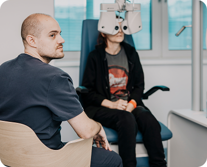
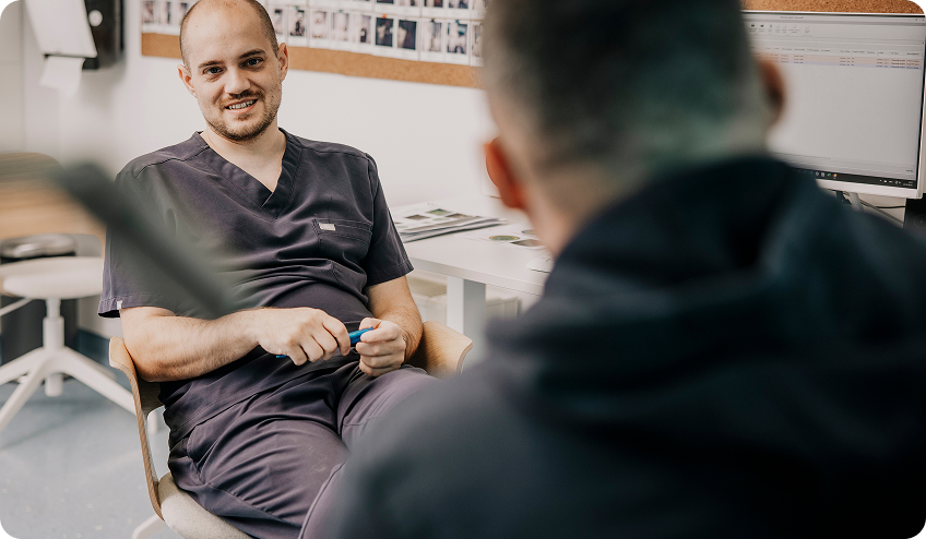
Doctor Ivan Gabrić with a patient
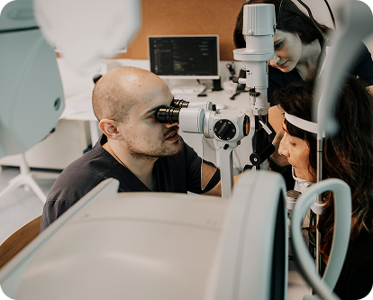
Ivan Gabrić, MD
The procedure is recommended for patients who meet the following criteria:
Corneal thickness greater than 390 microns
Clear cornea, without scars
Steeper corneal meridian up to a maximum of 58 diopters
Visual acuity with glasses 0.9 or lower
The procedure is not suitable for patients with other corneal diseases or for pregnant women.
How the Procedure is Performed
The procedure lasts about two hours and is performed under local anesthetic eye drops, completely painlessly. The main steps are:
- Removal of the corneal epithelial layer – the superficial layer is carefully removed with a fine spatula.
- Application of riboflavin (vitamin B2) – the cornea is soaked with concentrated riboflavin for 20-30 minutes. In addition to strengthening the cornea, riboflavin protects deeper eye structures from UV rays.
- UV lamp treatment – the cornea is treated with a UV lamp for 30 minutes with breaks every 5 minutes, during which additional Riboflavin is applied.
- Eye protection after the procedure – a soft contact lens is applied to the treated cornea and remains for 4-7 days. The patient continues with local corticosteroid eye drop therapy for the next two months.
On the first day after the procedure, severe discomfort or pain may be experienced, which patients are warned about in advance.
Recovery and Expected Results
The described procedure is based on the so-called Dresden protocol.
Actual results are evaluated after 6-12 months in adults, while in children, up to 24 months are needed for a final assessment.
After recovery, the patient can resume wearing contact lenses, often the same ones as before the procedure.
Cross-linking effectively stabilizes the cornea and slows the progression of keratoconus, thereby reducing the risk of needing a transplant in the future.
05
Cornea Lux
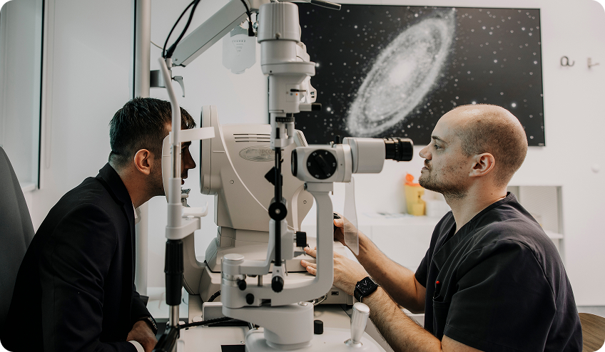
Cornea Lux is a new, tailor-made service from Svjetlost Eye Clinic, fully adapted to the individual needs of patients with corneal diseases.
It is intended for individuals with reduced visual acuity who, due to corneal changes, cannot function normally, for example, to obtain a driver’s license.
A personalized approach means that the diagnostic and treatment process is specifically tailored for each patient, as every scar or irregularity of the eye is unique.
Advantages over Classical Methods
The current generally accepted practice in Croatia and Southeast Europe is for patients with corneal diseases to undergo transplantation. Cornea Lux changes this approach. The primary goal of the service is to reduce the number of corneal surgeries and decrease the degree of disability in individuals with corneal diseases, including corneal degeneration (primarily keratoconus), granular dystrophy, and corneal injuries and infections.
Recovery is up to six times shorter than with classical transplantation, postoperative therapy is significantly shorter, and the need for immunosuppressive therapy is eliminated in many cases. Patients regain quality of vision and daily activities more quickly, and overall treatment costs and lost income due to sick leave are significantly reduced.
How Does Cornea Lux Work?
This innovative method combines known procedures and interventions in the treatment of corneal diseases, including:
Special diagnostic procedures
Special medication regimen before and after the procedure
Personalized ablation profiles
Advanced models of tissue immune modulation after the procedure
The procedure lasts 30 to 45 minutes, which is slightly longer than standard laser vision correction (about 20 minutes), but it offers multiple advantages in terms of safety, precision, and long-term effect.
What Does Cornea Lux Replace or Improve?
There are some alternatives to corneal transplantation on the market, but none are fully personalized:

Using the PTK module on the laser
Tissue is removed uniformly from the entire corneal surface, without precise targeting of scars or irregularities.

Manual scraping of dystrophy
Manual, less precise than a programmed laser.

Topography-guided keratoconus treatment
Marginally accepted, often avoided due to fear of corneal destabilization.
Cornea Lux integrates all these approaches into a unique protocol, personalized for each patient, reducing the need for classical transplantation and improving overall visual quality.
06
CAIRS
CAIRS (Corneal Allogenic Intrastromal Ring Segments) is one of the most advanced contemporary methods for treating keratoconus and other corneal ectasias. For many patients, CAIRS represents a new option between conservative treatment and corneal transplantation.
What is CAIRS?
CAIRS is a modern surgical method in which specially shaped segments made from donor human cornea are implanted into the cornea. The segments are placed in the middle layers of the cornea where they provide mechanical support to the weakened tissue, help remodel its shape, and reduce the deformation characteristic of keratoconus.
The goals of the procedure are:
Improve the cornea
Reduce irregular astigmatism
Improve visual quality
Increase contact lens tolerance
Delay or avoid the need for corneal transplantation
Why is CAIRS Different?
Traditional intrastromal rings are made of synthetic materials.
CAIRS uses biological corneal tissue, which allows for more natural integration and biomechanical compatibility with the eye.
Advantages of the CAIRS method include:
- biological implant made from donor cornea
- more natural integration with eye tissue
- stabilization and remodeling of the cornea
- reduction of corneal surface irregularities
- improvement of visual quality
- possibility of combining with corneal cross-linking procedure
- potential reduction in the need for corneal transplantation
What does the procedure involve?
The procedure is performed using a femtosecond laser, which allows for exceptional precision.
During the procedure:
- A tunnel is created within the cornea using a femtosecond laser.
- One or two CAIRS segments are implanted into the prepared space.
- If necessary, the procedure is combined with the corneal cross-linking method for additional corneal stabilization.
The procedure is minimally invasive, and the patient returns to daily activities soon after the procedure, as recommended by the doctor.
Who is a Candidate for CAIRS?
CAIRS may be a suitable solution for patients who:
Have keratoconus or another corneal ectasia
No longer achieve satisfactory vision with glasses
Have difficulty tolerating contact lenses
Have progressive corneal deformation
Wish to delay or avoid corneal transplantation
What Results Can Be Expected?
Clinical experience shows that CAIRS can lead to:
Improvement in visual acuity
Reduction of irregular astigmatism
Improvement in corneal topography
Better corneal stability
Higher quality of daily vision
The results develop gradually over months after the procedure, as the cornea undergoes a process of adaptation and stabilization.
CAIRS at Svjetlost
At Svjetlost Special Hospital for Ophthalmology, we apply the most modern methods for treating corneal diseases, including CAIRS technology for carefully selected keratoconus patients. Each patient undergoes a detailed diagnostic evaluation to choose the optimal treatment and achieve the best possible functional outcome. Our goal is not only to improve vision but also to preserve corneal health and patients’ quality of life in the long term.
07
Your medical team – our physicians


Member of the Management Board

Ophthalmologist

Ophthalmologist

Ophthalmologist
08