Blog
Keratoconus no longer means an inevitable corneal transplant
What is keratoconus?
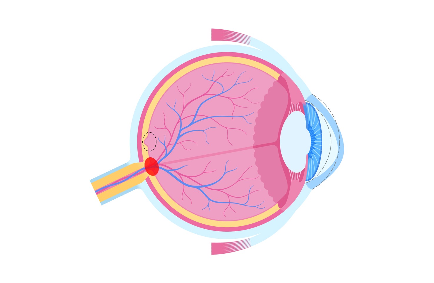
Keratoconus is a progressive, non-inflammatory corneal disease in which the cornea, the clear front part of the eye, gradually thins and bulges into a cone shape. Because the cornea provides most of the eye’s refractive power, even small changes in its shape can significantly distort vision and cause irregular astigmatism.
The condition usually appears in the teenage years or the twenties. It progresses fastest in young adulthood and generally slows down with age. Early signs include a frequently changing glasses prescription, double images, glare around lights, and vision that can no longer be fully corrected with glasses.
Who is at higher risk?
Estimates vary, but keratoconus affects at least one in two thousand people, and thanks to improved diagnostics it is now detected earlier than ever. It is associated with a family history of the disease, early onset, and eye rubbing, particularly in people with allergies or atopic dermatitis.
How is it diagnosed?
Keratoconus is diagnosed with corneal topography or tomography, a painless, non-contact scan that produces a detailed map of the cornea’s curvature and thickness. Repeat scans are used to monitor whether the disease is progressing.
Step-by-step treatment
First and foremost, stop rubbing your eyes. Eye rubbing is the most important modifiable risk factor for progression, and avoiding it is the foundation of every other step of treatment.
1. CAIRS – reshaping the cornea
CAIRS (Corneal Allogenic Intrastromal Ring Segments) is one of the most advanced modern treatments for keratoconus and other corneal ectasias. In this surgical procedure, specially shaped segments of donor human corneal tissue are implanted into the corneal stroma. There they provide structural support to the weakened tissue, help remodel the cornea and reduce the irregularity typical of keratoconus.
What does the procedure involve?
The procedure is performed with a femtosecond laser, which allows exceptional precision:
- The femtosecond laser creates an intrastromal channel within the cornea.
- One or two CAIRS segments are implanted into the channel.
- If needed, the procedure is combined with corneal cross-linking to further stabilise the cornea.
The procedure is minimally invasive, and patients can soon return to their daily activities, as advised by their ophthalmologist.
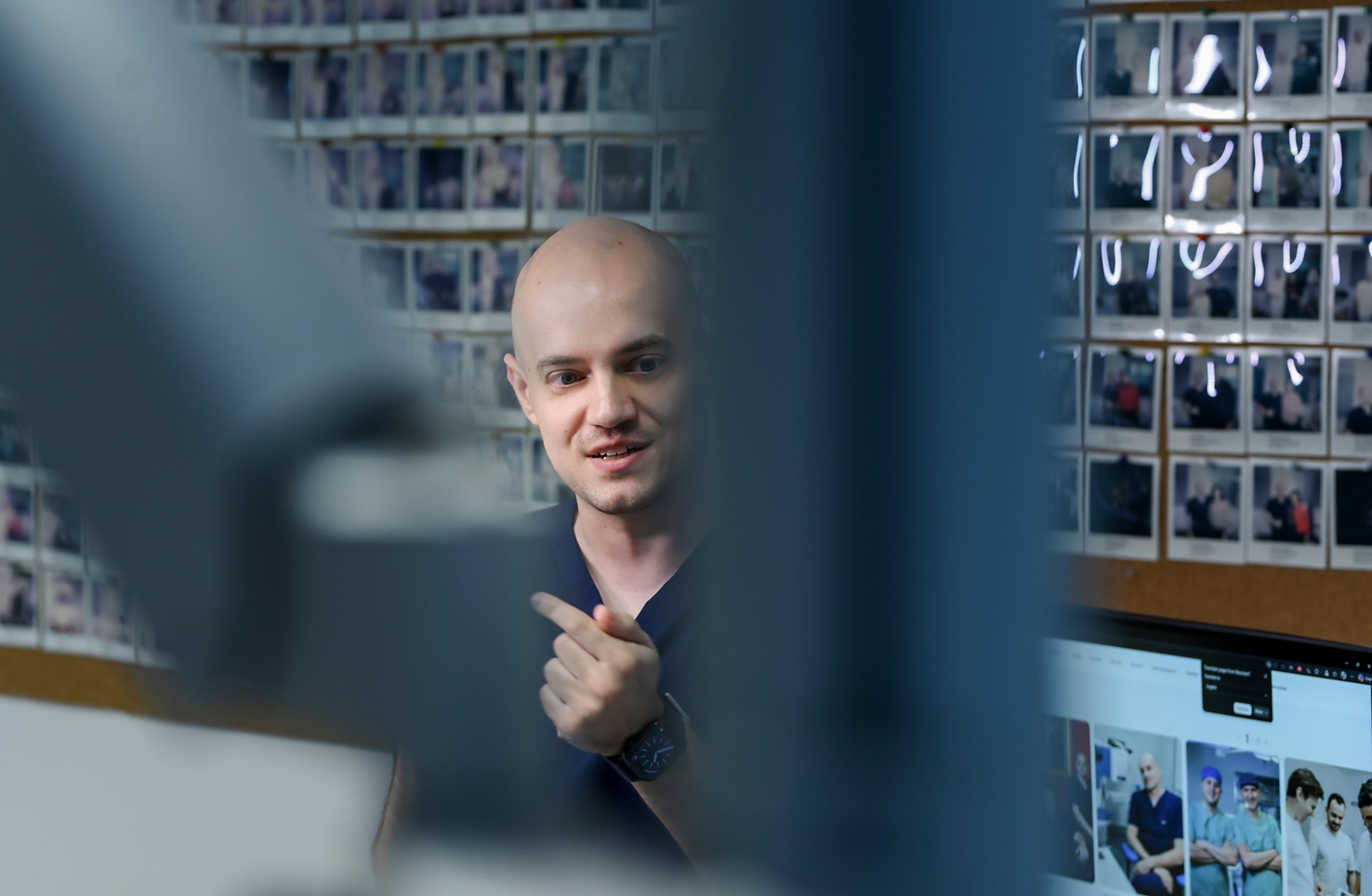
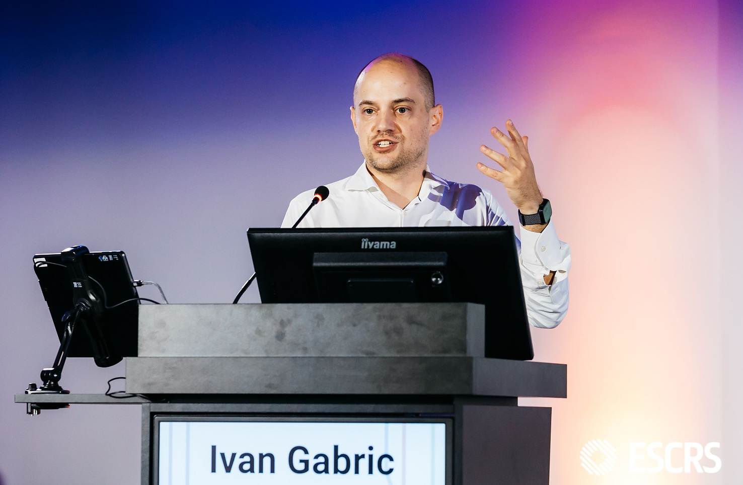
The first CAIRS procedures in Croatia were performed by ophthalmologist Dr Ivan Gabrić at Svjetlost Eye Clinic.
2. Corneal cross-linking – stabilising the cornea
Corneal cross-linking strengthens the weakened collagen bonds in the cornea and slows the progression of keratoconus. It is recommended when the disease is progressing and can be performed together with CAIRS or as a separate procedure.
The procedure takes about two hours and is performed under topical anaesthesia (anaesthetic eye drops), so the procedure itself is painless. The main steps are:
- Epithelial removal (epi-off) – the surface layer of the cornea is carefully removed with a fine spatula.
- Riboflavin application – the cornea is soaked with riboflavin (vitamin B2) for 20 to 30 minutes. Riboflavin enables the cross-linking reaction and protects the deeper structures of the eye from UV exposure.
- UVA irradiation – the cornea is exposed to UVA light for 30 minutes, with pauses every 5 minutes to apply additional riboflavin.
- Post-operative care – a bandage contact lens is placed on the cornea for 4 to 7 days, and the patient uses topical corticosteroid drops for the following two months.
3. Correcting residual refractive error
Residual refractive error is corrected with glasses or contact lenses. For higher prescriptions, an implantable phakic lens (ICL) may be an option, and laser vision correction may be considered in selected cases.
4. Corneal transplant (keratoplasty) – only when necessary
A corneal transplant is reserved for the small number of patients whose disease continues to progress despite all of the above treatments.
Is your prescription changing, or are your glasses no longer giving you clear vision?
The sooner keratoconus is diagnosed, the easier it is to treat. Book a corneal examination at Svjetlost Eye Clinic, where our specialists will recommend a treatment tailored to your eyes.
Istražite naše novosti i stručne blogove
News & Expert advice
-
![]()
News
EASE – a new surface laser vision correction method at Svjetlost Eye Clinic
Svjetlost Eye Clinic now offers its patients EASE, the newest method of laser vision correction. EASE (Epithelial All-femtosecond Surface Excision) was developed by the German company SCHWIND eye-tech-solutions and is performed with the SCHWIND ATOS® laser.
-
![]()
News
Svjetlost Eye Clinic launches the international STELLAR clinical study
Svjetlost Eye Clinic is the sponsor of STELLAR, an international clinical study that will systematically follow the outcomes of transepithelial lenticule removal (TLR). TLR is a new laser method for correcting myopia and astigmatism, also known by its commercial name, EASE.
-
![]()
News
Svjetlost Eye Clinic at the 44th ESCRS Congress
Leading world ophthalmologists in London.