Blog
What does an ophthalmological exam look like?
Let's find out what happens behind those blue doors after our name is called.
Fear of white coats or examinations should not prevent anyone from checking their health, including their eye health.
The answer to the question of what awaits us behind the closed doors of our doctors’ clinics can help dispel fears and allow us to trust and entrust our problems to the Svjetlost Eye Clinic.
So let’s find out what happens behind those blue doors after our name is called.
Ophthalmological examinations are completely painless , we like to say that we look at lights, colors, letters and numbers and that’s it, the examination is already over.
Each examination begins with introductions and conversation . Through mutual communication, we learn what is troubling the person who comes, and through more detailed questions, we discern what the problem is.
The doctor will also ask you about your general health, whether you are taking any medications, whether you have any known allergies, and whether there has been any eye disease in your family. If you wear glasses, bring them with you to the examination, as well as any previous findings if you have them.
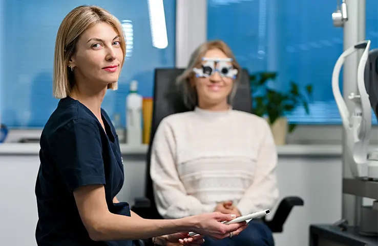
After a conversation and an assessment of what is happening with the patient, an examination usually begins with an eye test.
The patient is put on trial glasses, and visual acuity is checked and the diopter is determined , first in one eye, then in the other, for distance and near. The exact diopter is reached, so you should not be confused if the image is not immediately clear at the beginning of the vision test. When your doctor changes your glasses, try to distinguish which one has clearer letters and sharper edges to help him reach the correct diopter.
After that, the eye pressure is checked, most often with a small device that measures it in a split second, painlessly and without discomfort for the patient.
The examination with a biomicroscope is simple, the patient places their chin in the bowl and rests their forehead on a special strip. The eye is viewed under different lights and magnifications to see all the details, and during this time the patient sees light shining on it from different angles.
This is followed by pupil dilation . Don’t worry, we don’t have any scary devices to do this, but rather the pupils are dilated very elegantly by instilling drops, and it doesn’t hurt at all. The duration of pupil dilation varies depending on whether it’s a child or an adult and whether the goal of pupil dilation is to completely relax the muscles with which we can hide our diopter or whether we just need a wider field of view inside the eye to assess the condition of the lens and the fundus .
The patient is then placed back in the waiting room while the pupils dilate, which takes about 30 minutes on average. Due to the dilation of the pupils, the patient’s vision will be blurred, especially at close range, for 2-3 hours after the examination.
Once the pupils have dilated, the patient is called back to the clinic and we continue with the second part of the examination. This consists of an examination of the fundus and possibly re-measuring the diopter if the doctor deems it necessary.
During the examination of the fundus, the patient is again positioned under the biomicroscope and asked to look straight ahead. A small magnifying glass is held in front of the patient’s eye, which does not touch the eye, giving the doctor enough magnification to see inside the eye, the optic nerve, macula, blood vessels, and retina. During the examination, the doctor tells the patient which direction to look in order to examine the entire inside of the eye.
By looking into the posterior segment of the eye, we can also assess the condition of the blood vessels, which are a mirror of the vessels in our entire body, which is especially important in diseases such as diabetes and blood pressure. It is important that the patient does not close the other eye, but actively looks with it and concentrates on it in order to reduce the glare of light on the eye being examined as much as possible.
After the examination is complete, the doctor explains to the patient what he saw during the examination and together they agree on further steps in treatment, if necessary, and follow-up examinations.
Istražite naše novosti i stručne blogove
News & Expert advice
-
![]()
News
Svjetlost Eye Clinic Publishes the First Croatian Textbook on Refractive Surgery
On 20 November, the first day of the 16th Congress of the Croatian Society for Cataract and Refractive Surgery (CSCRS), “New Trends in Ophthalmology”, Svjetlost Eye Clinic will publish the textbook Refrakcijska kirurgija (Refractive Surgery).
-
![]()
News
Dr. Ivan Gabrić Speaks at SCHWIND DACH Meeting of Ophthalmologists from Germany, Austria and Switzerland
Dr. Ivan Gabrić of Svjetlost Eye Clinic was one of the speakers at the SCHWIND DACH User Meeting, a professional gathering of ophthalmologists from Germany, Austria and Switzerland who work with lasers made by the German company SCHWIND.
-
![]()
News
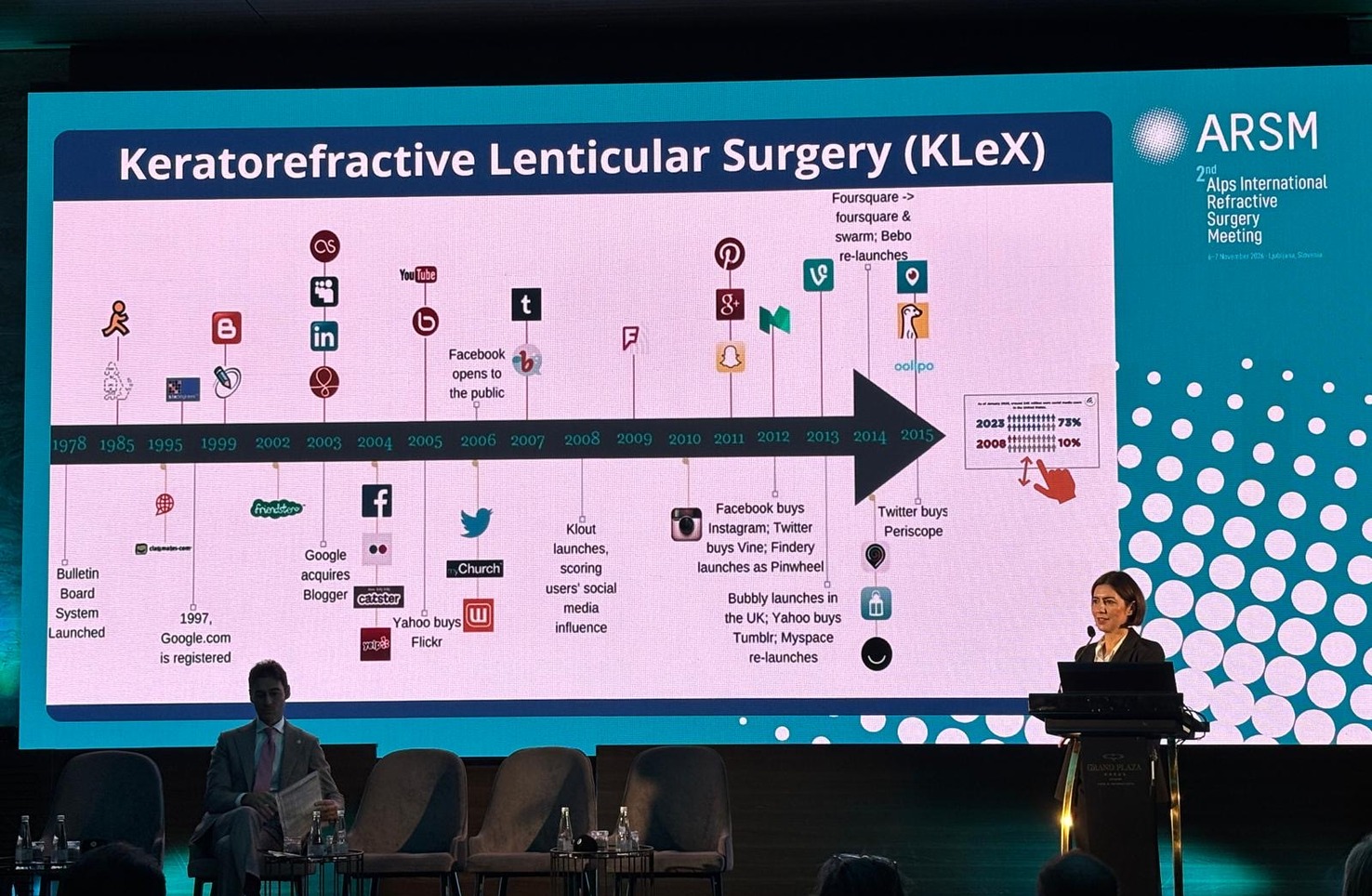
Prof. Maja Bohač at the 2nd ARSM in Ljubljana
Prof. Maja Bohač, MD, PhD, Head of the Department of Refractive Surgery at Svjetlost Eye Clinic, gave an invited lecture titled “Current State of Lenticular Extraction” at the 2nd Alps Refractive Surgery Meeting (ARSM).